Krestel Heinz, Weisstanner Christian, Hess Christian W, Bassetti Claudio L, Nirkko Arto, Wiest Roland
Department of Neurology, Inselspital, Bern University Hospital, University of Bern, Freiburgstrasse 10, 3010, Bern, Switzerland,
Brain Struct Funct. 2015 Mar;220(2):803-12. doi: 10.1007/s00429-013-0684-6. Epub 2013 Dec 12.
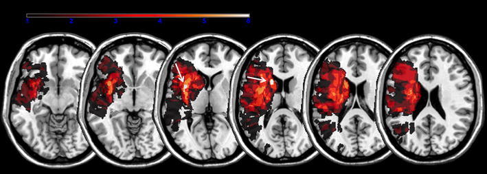
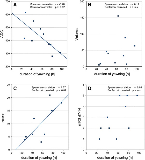
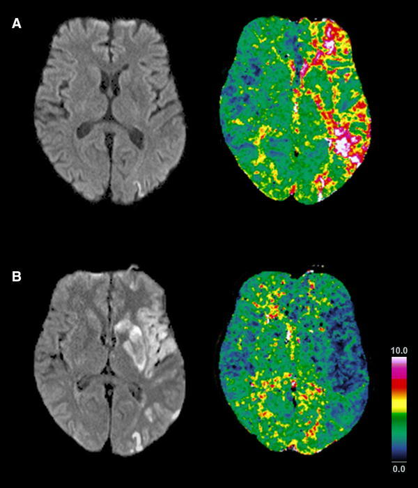
Abnormal yawning is an underappreciated phenomenon in patients with ischemic stroke. We aimed at identifying frequently affected core regions in the supratentorial brain of stroke patients with abnormal yawning and contributing to the anatomical network concept of yawning control. Ten patients with acute anterior circulation stroke and ≥3 yawns/15 min without obvious cause were analyzed. The NIH stroke scale (NIHSS), Glasgow Coma Scale (GCS), symptom onset, period with abnormal yawning, blood oxygen saturation, glucose, body temperature, blood pressure, heart rate, and modified Rankin scale (mRS) were assessed for all patients. MRI lesion maps were segmented on diffusion-weighted images, spatially normalized, and the extent of overlap between the different stroke patterns was determined. Correlations between the period with abnormal yawning and the apparent diffusion coefficient (ADC) in the overlapping regions, total stroke volume, NIHSS and mRS were performed. Periods in which patients presented with episodes of abnormal yawning lasted on average for 58 h. Average GCS, NIHSS, and mRS scores were 12.6, 11.6, and 3.5, respectively. Clinical parameters were within normal limits. Ischemic brain lesions overlapped in nine out of ten patients: in seven patients in the insula and in seven in the caudate nucleus. The decrease of the ADC within the lesions correlated with the period with abnormal yawing (r = -0.76, Bonferroni-corrected p = 0.02). The stroke lesion intensity of the common overlapping regions in the insula and the caudate nucleus correlates with the period with abnormal yawning. The insula might be the long sought-after brain region for serotonin-mediated yawning.
异常打哈欠是缺血性中风患者中一种未得到充分认识的现象。我们旨在确定有异常打哈欠的中风患者幕上脑内经常受影响的核心区域,并为打哈欠控制的解剖网络概念提供依据。分析了10例急性前循环中风且无明显原因打哈欠≥3次/15分钟的患者。对所有患者评估了美国国立卫生研究院卒中量表(NIHSS)、格拉斯哥昏迷量表(GCS)、症状发作时间、异常打哈欠期、血氧饱和度、血糖、体温、血压、心率和改良Rankin量表(mRS)。在扩散加权图像上对MRI病变图进行分割、空间归一化,并确定不同中风模式之间的重叠程度。对重叠区域内异常打哈欠期与表观扩散系数(ADC)、总中风体积、NIHSS和mRS之间进行相关性分析。患者出现异常打哈欠发作的时间平均持续58小时。平均GCS、NIHSS和mRS评分分别为12.6、11.6和3.5。临床参数在正常范围内。10例患者中有9例的缺血性脑病变有重叠:7例在岛叶,7例在尾状核。病变内ADC的降低与异常打哈欠期相关(r = -0.76,经Bonferroni校正的p = 0.02)。岛叶和尾状核共同重叠区域的中风病变强度与异常打哈欠期相关。岛叶可能是长期以来寻找的5-羟色胺介导打哈欠的脑区。