State Key Laboratory of Cellular Stress Biology, Innovation Center for Cell Biology, School of Life Sciences, Xiamen University, Xiamen, Fujian 361005, China.
Cell Res. 2014 Jan;24(1):105-21. doi: 10.1038/cr.2013.171. Epub 2013 Dec 24.
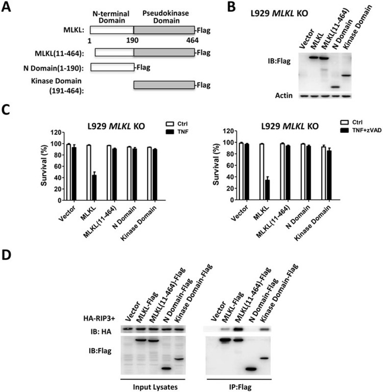
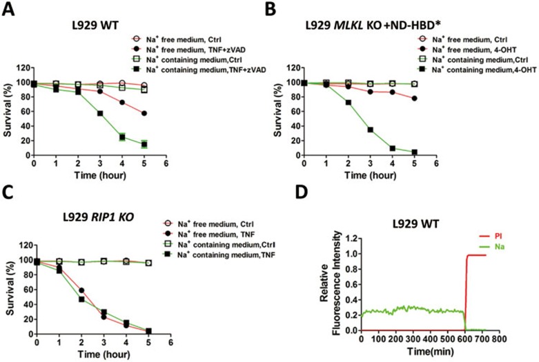
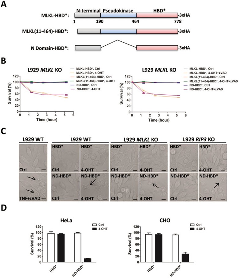
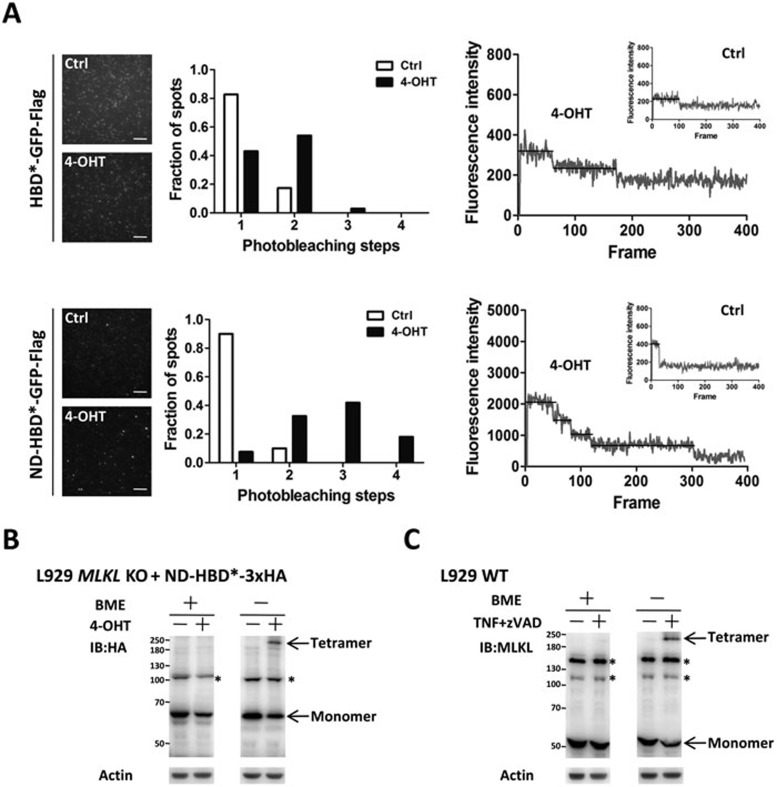
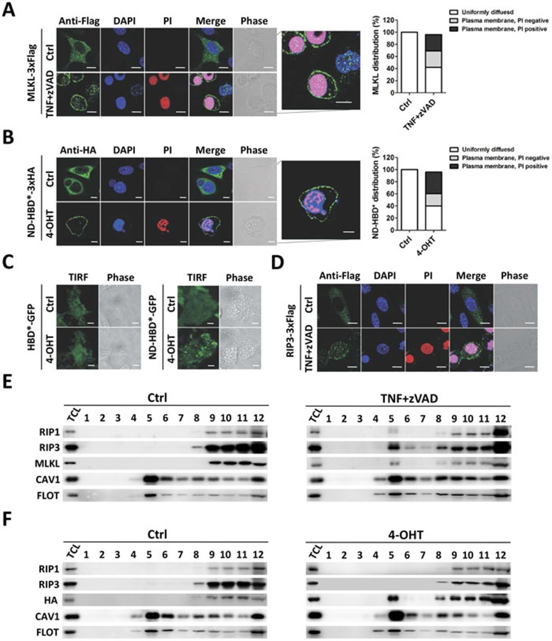
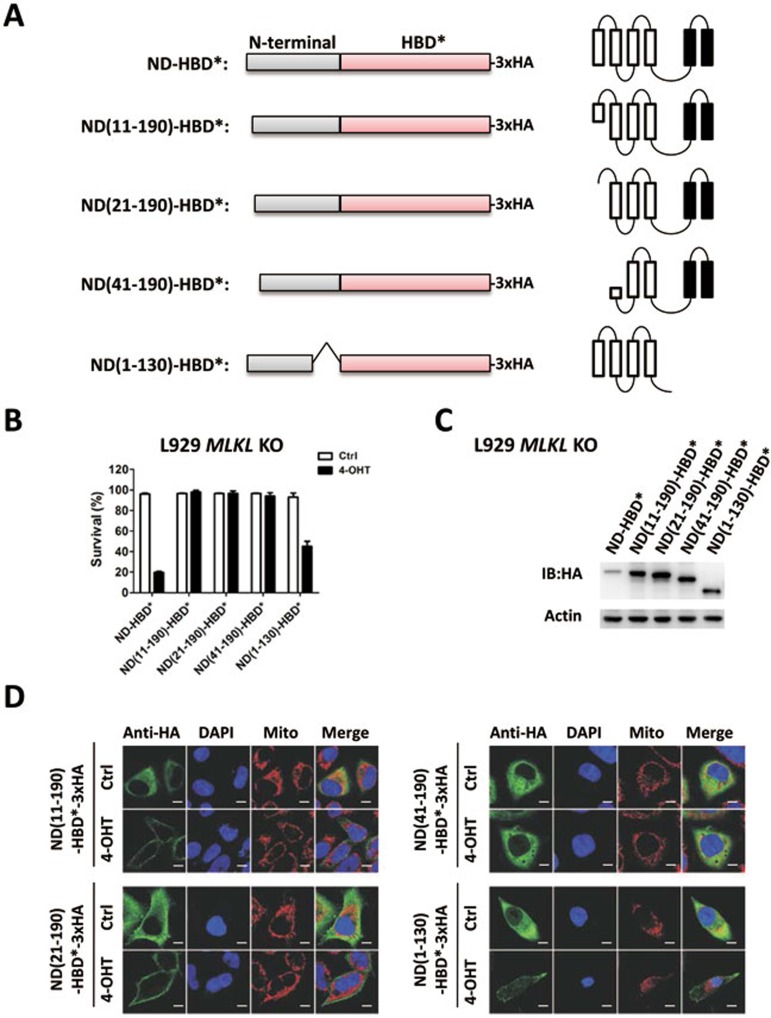
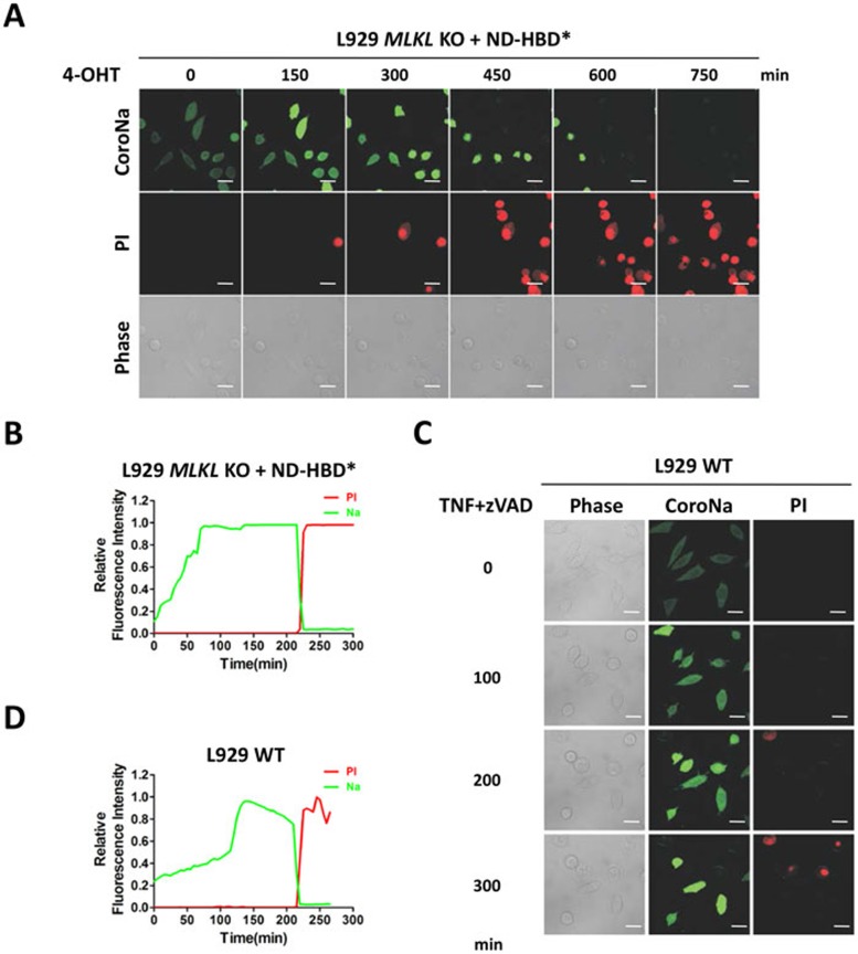
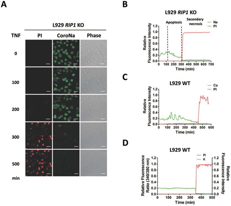
Mixed lineage kinase domain-like protein (MLKL) was identified to function downstream of receptor interacting protein 3 (RIP3) in tumor necrosis factor-α (TNF)-induced necrosis (also called necroptosis). However, how MLKL functions to mediate necroptosis is unknown. By reconstitution of MLKL function in MLKL-knockout cells, we showed that the N-terminus of MLKL is required for its function in necroptosis. The oligomerization of MLKL in TNF-treated cells is essential for necroptosis, as artificially forcing MLKL together by using the hormone-binding domain (HBD*) triggers necroptosis. Notably, forcing together the N-terminal domain (ND) but not the C-terminal kinase domain of MLKL causes necroptosis. Further deletion analysis showed that the four-α-helix bundle of MLKL (1-130 amino acids) is sufficient to trigger necroptosis. Both the HBD*-mediated and TNF-induced complexes of MLKL(ND) or MLKL are tetramers, and translocation of these complexes to lipid rafts of the plasma membrane precedes cell death. The homo-oligomerization is required for MLKL translocation and the signal sequence for plasma membrane location is located in the junction of the first and second α-helices of MLKL. The plasma membrane translocation of MLKL or MLKL(ND) leads to sodium influx, and depletion of sodium from the cell culture medium inhibits necroptosis. All of the above phenomena were not seen in apoptosis. Thus, the MLKL oligomerization leads to translocation of MLKL to lipid rafts of plasma membrane, and the plasma membrane MLKL complex acts either by itself or via other proteins to increase the sodium influx, which increases osmotic pressure, eventually leading to membrane rupture.
混合谱系激酶结构域样蛋白(MLKL)被鉴定为在肿瘤坏死因子-α(TNF)诱导的坏死(也称为坏死性凋亡)中作为受体相互作用蛋白 3(RIP3)的下游发挥作用。然而,MLKL 如何发挥作用介导坏死性凋亡尚不清楚。通过在 MLKL 敲除细胞中重建 MLKL 功能,我们表明 MLKL 的 N 端对于其在坏死性凋亡中的功能是必需的。TNF 处理细胞中 MLKL 的寡聚化对于坏死性凋亡是必不可少的,因为通过使用激素结合结构域(HBD*)人为地迫使 MLKL 聚集会引发坏死性凋亡。值得注意的是,迫使 MLKL 的 N 端结构域(ND)而不是 C 端激酶结构域聚集会导致坏死性凋亡。进一步的缺失分析表明,MLKL(1-130 个氨基酸)的四-α-螺旋束足以引发坏死性凋亡。HBD*-介导的和 TNF 诱导的 MLKL(ND)或 MLKL 复合物都是四聚体,并且这些复合物向质膜脂筏的易位先于细胞死亡。同源寡聚化是 MLKL 易位所必需的,并且质膜位置的信号序列位于 MLKL 的第一个和第二个α-螺旋的连接处。MLKL 或 MLKL(ND)的质膜易位导致钠离子内流,并且从细胞培养基中耗尽钠离子可抑制坏死性凋亡。在凋亡中没有观察到所有这些现象。因此,MLKL 寡聚化导致 MLKL 向质膜脂筏的易位,并且质膜 MLKL 复合物通过自身或通过其他蛋白增加钠离子内流,从而增加渗透压,最终导致膜破裂。