• 文献检索
  • 文档翻译
  • 深度研究
  • 学术资讯
  • Suppr Zotero 插件Zotero 插件
  • 邀请有礼
  • 套餐&价格
  • 历史记录
应用&插件
Suppr Zotero 插件Zotero 插件浏览器插件Mac 客户端Windows 客户端微信小程序
定价
高级版会员购买积分包购买API积分包
服务
文献检索文档翻译深度研究API 文档MCP 服务
关于我们
关于 Suppr公司介绍联系我们用户协议隐私条款
关注我们

Suppr 超能文献

核心技术专利:CN118964589B侵权必究
粤ICP备2023148730 号-1Suppr @ 2026

文献检索

告别复杂PubMed语法,用中文像聊天一样搜索,搜遍4000万医学文献。AI智能推荐,让科研检索更轻松。

立即免费搜索

文件翻译

保留排版,准确专业,支持PDF/Word/PPT等文件格式,支持 12+语言互译。

免费翻译文档

深度研究

AI帮你快速写综述,25分钟生成高质量综述,智能提取关键信息,辅助科研写作。

立即免费体验

应用对比增强超声技术研究一组疑似正中神经单一神经病变的食蟹猴的神经内血管分布。

Use of contrast-enhanced sonography to investigate intraneural vascularity in a cohort of Macaca fascicularis with suspected median mononeuropathy.

机构信息

RT, RVS, The Ohio State University, 453 W 10th Ave, Columbus, OH 43210 USA.

出版信息

J Ultrasound Med. 2014 Jan;33(1):103-9. doi: 10.7863/ultra.33.1.103.

DOI:10.7863/ultra.33.1.103
PMID:24371104
原文链接:https://pmc.ncbi.nlm.nih.gov/articles/PMC4040227/
Abstract

OBJECTIVES

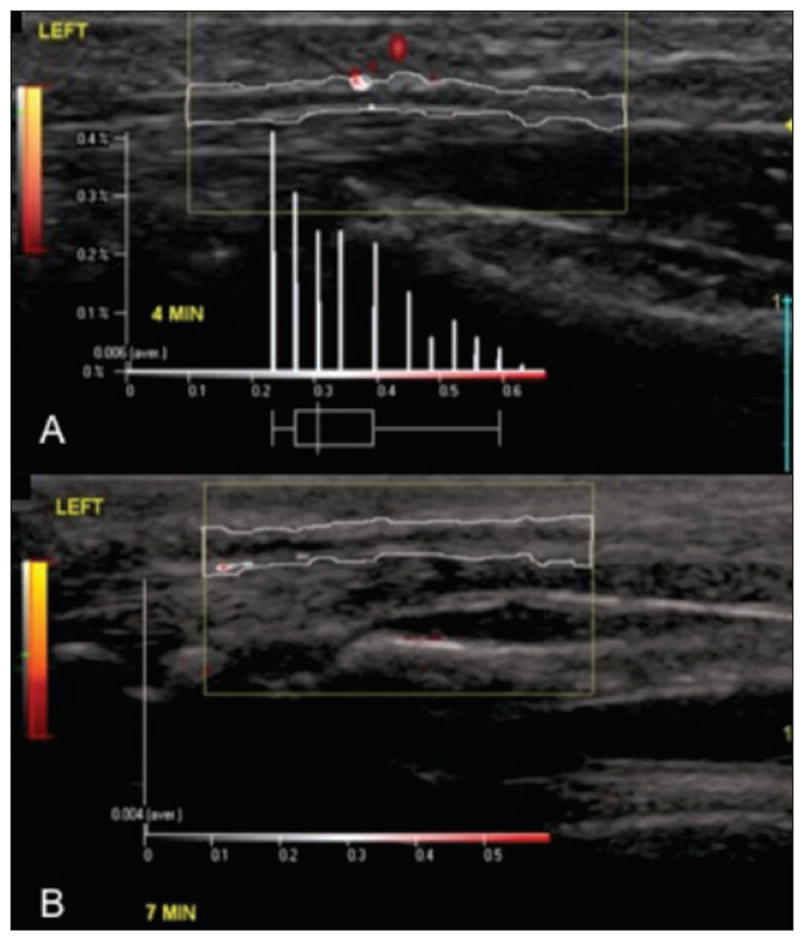
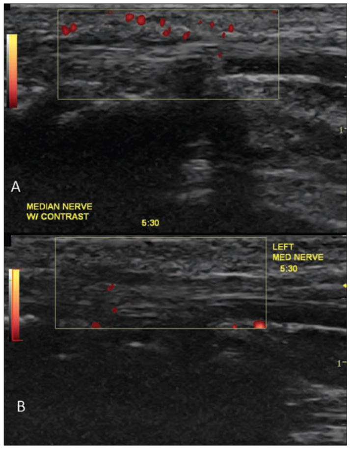
The purpose of this study was to provide clinical evidence of the use of contrast-enhanced sonography in detecting and quantifying changes in intraneural vascularity due to median mononeuropathy.

METHODS

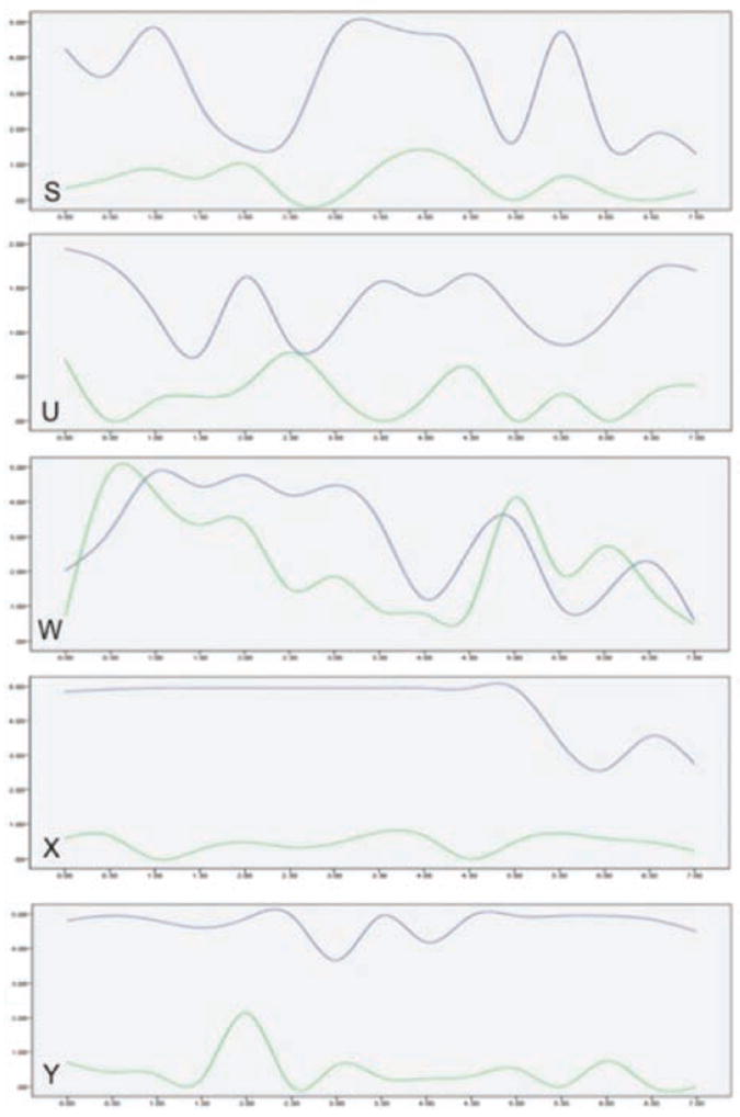
Five Macaca fascicularis monkeys were exposed to 20 weeks of repetitive work to increase their risk of developing median mononeuropathy. Contrast-enhanced sonograms were obtained in 30-second increments for 7 minutes while a contrast agent was being delivered. Data were collected immediately at the conclusion of the 20-week work exposure and then again during a recovery phase approximately 3 months after the completion of work. Quantitative analysis and trend graphs were used to analyze median nerve perfusion intensity. This study also compared the use of both manual counting of pixels and semiautomatic measurement using specialized software.

RESULTS

Based on the average data, maximum intensity values were identified as the best indicators of nerve hyperemia. Paired t tests demonstrated significantly higher maximum intensities in the working stage for 4 of the 5 subjects (P < .01).

CONCLUSIONS

This study provides preliminary evidence that (1) in a controlled exposure model, a change in intraneural vascularity of the median nerve between working and recovery can be observed; (2) this vascular change can be measured using an objective technique that quantifies the intensity of vascularity; and (3) contrast-enhanced sonography may improve the ability to reliably capture and measure low-flow microvascularity.

摘要

目的

本研究旨在提供临床证据,证明对比增强超声在检测和量化正中神经单神经病引起的神经内血管变化方面的作用。

方法

将 5 只食蟹猴暴露于 20 周的重复性工作中,以增加其发生正中神经单神经病的风险。在注射对比剂的同时,以 30 秒为间隔进行 7 分钟的对比增强超声检查。在 20 周工作暴露结束时立即收集数据,然后在工作完成约 3 个月后的恢复期再次收集数据。使用定量分析和趋势图分析正中神经灌注强度。本研究还比较了手动计数像素和使用专用软件进行半自动测量的两种方法。

结果

根据平均数据,最大强度值被确定为神经充血的最佳指标。配对 t 检验显示,5 名受试者中有 4 名在工作阶段的最大强度显著升高(P <.01)。

结论

本研究初步证明:(1)在受控暴露模型中,可以观察到正中神经内神经血管变化在工作和恢复之间的变化;(2)可以使用客观技术测量这种血管变化,该技术可以量化血管密度;(3)对比增强超声可能提高可靠捕获和测量低流速微血管的能力。

https://cdn.ncbi.nlm.nih.gov/pmc/blobs/838a/4040227/a44f4e723b5c/nihms569795f3.jpg
https://cdn.ncbi.nlm.nih.gov/pmc/blobs/838a/4040227/26ad36c0633f/nihms569795f1.jpg
https://cdn.ncbi.nlm.nih.gov/pmc/blobs/838a/4040227/2c08dfe1e0be/nihms569795f2.jpg
https://cdn.ncbi.nlm.nih.gov/pmc/blobs/838a/4040227/a44f4e723b5c/nihms569795f3.jpg
https://cdn.ncbi.nlm.nih.gov/pmc/blobs/838a/4040227/26ad36c0633f/nihms569795f1.jpg
https://cdn.ncbi.nlm.nih.gov/pmc/blobs/838a/4040227/2c08dfe1e0be/nihms569795f2.jpg
https://cdn.ncbi.nlm.nih.gov/pmc/blobs/838a/4040227/a44f4e723b5c/nihms569795f3.jpg

相似文献

1
Use of contrast-enhanced sonography to investigate intraneural vascularity in a cohort of Macaca fascicularis with suspected median mononeuropathy.应用对比增强超声技术研究一组疑似正中神经单一神经病变的食蟹猴的神经内血管分布。
J Ultrasound Med. 2014 Jan;33(1):103-9. doi: 10.7863/ultra.33.1.103.
2
Detection of Intraneural Median Nerve Microvascularity Using Contrast-Enhanced Sonography: A Pilot Study.使用超声造影检测神经内正中神经微血管:一项初步研究。
J Ultrasound Med. 2016 Jun;35(6):1309-16. doi: 10.7863/ultra.15.07012. Epub 2016 May 5.
3
Color and power Doppler US for diagnosing carpal tunnel syndrome and determining its severity: a quantitative image processing method.彩色和能量多普勒超声用于诊断腕管综合征及其严重程度的定量图像处理方法。
Radiology. 2011 Nov;261(2):499-506. doi: 10.1148/radiol.11110150. Epub 2011 Sep 7.
4
Reliability of peripheral intraneural microhemodynamics evaluation by using contrast-enhanced ultrasonography.使用超声造影评估周围神经内微血流动力学的可靠性。
J Med Ultrason (2001). 2014 Oct;41(4):481-6. doi: 10.1007/s10396-014-0533-0. Epub 2014 Mar 19.
5
Longitudinal design for sonographic measurement of median nerve swelling with controlled exposure to physical work using an animal model.使用动物模型对超声测量正中神经肿胀的纵向设计,该模型通过控制物理工作的暴露程度来实现。
Ultrasound Med Biol. 2013 Dec;39(12):2492-7. doi: 10.1016/j.ultrasmedbio.2013.08.008. Epub 2013 Oct 16.
6
Intraneural microvascular patterns of the median nerve assessed using contrast-enhanced ultrasonography in carpal tunnel syndrome.在腕管综合征中使用超声造影评估正中神经的神经内微血管模式。
J Hand Surg Eur Vol. 2016 Feb;41(2):229-31. doi: 10.1177/1753193415570222. Epub 2015 Feb 10.
7
Ultrasound for diagnosis of carpal tunnel syndrome: comparison of different methods to determine median nerve volume and value of power Doppler sonography.超声诊断腕管综合征:不同方法测量正中神经体积的比较及能量多普勒超声的价值。
Ann Rheum Dis. 2013 Dec;72(12):1934-9. doi: 10.1136/annrheumdis-2012-202328. Epub 2012 Dec 4.
8
Relationship between intraneural vascular flow measured with sonography and carpal tunnel syndrome diagnosis based on electrodiagnostic testing.超声测量神经内血流与基于电诊断检测的腕管综合征诊断的关系。
J Ultrasound Med. 2012 May;31(5):729-36. doi: 10.7863/jum.2012.31.5.729.
9
Superb Microvascular Imaging of the Median Nerve in Carpal Tunnel Syndrome: An Electrodiagnostic and Ultrasonographic Study.腕管综合征正中神经的超微血管成像:一项电诊断和超声研究
J Ultrasound Med. 2018 Dec;37(12):2855-2861. doi: 10.1002/jum.14645. Epub 2018 Apr 16.
10
Blood Flow Changes in Subsynovial Connective Tissue on Contrast-Enhanced Ultrasonography in Patients With Carpal Tunnel Syndrome Before and After Surgical Decompression.腕管综合征患者手术减压前后,对比增强超声检查下腕关节滑膜下结缔组织血流变化
J Ultrasound Med. 2018 Jul;37(7):1597-1604. doi: 10.1002/jum.14500. Epub 2017 Nov 24.

引用本文的文献

1
Peripheral nerve ultrasound: a survival guide for the practicing radiologist with updates.外周神经超声:执业放射科医生生存指南及更新内容
Ultrasound J. 2025 Mar 26;17(1):21. doi: 10.1186/s13089-024-00387-0.
2
Assessing the Potential for Error in Investigating Intraneural Vascularity: A Need for a Standardized Imaging Protocol.评估神经内血管研究中潜在的误差:对标准化成像方案的需求。
J Diagn Med Sonogr. 2023 Nov;39(6):549-559. doi: 10.1177/87564793231193396.
3
Value of superb microvascular imaging ultrasonography in the diagnosis of carpal tunnel syndrome: Compared with color Doppler and power Doppler.超微血管成像超声检查在腕管综合征诊断中的价值:与彩色多普勒和能量多普勒的比较
Medicine (Baltimore). 2017 May;96(21):e6862. doi: 10.1097/MD.0000000000006862.
4
Carpal tunnel syndrome severity staging using sonographic and clinical measures.使用超声和临床测量对腕管综合征进行严重程度分期
Muscle Nerve. 2015 Jun;51(6):838-45. doi: 10.1002/mus.24478. Epub 2015 Apr 24.

本文引用的文献

1
Morphologic Characterization of Intraneural Flow Associated With Median Nerve Pathology.与正中神经病变相关的神经内血流的形态学特征
J Diagn Med Sonogr. 2012 Jan;28(1):11-19. doi: 10.1177/8756479311426777.
2
Relationship between intraneural vascular flow measured with sonography and carpal tunnel syndrome diagnosis based on electrodiagnostic testing.超声测量神经内血流与基于电诊断检测的腕管综合征诊断的关系。
J Ultrasound Med. 2012 May;31(5):729-36. doi: 10.7863/jum.2012.31.5.729.
3
Color and power Doppler US for diagnosing carpal tunnel syndrome and determining its severity: a quantitative image processing method.彩色和能量多普勒超声用于诊断腕管综合征及其严重程度的定量图像处理方法。
Radiology. 2011 Nov;261(2):499-506. doi: 10.1148/radiol.11110150. Epub 2011 Sep 7.
4
Quantitative assessment of urethral vascularity in nulliparous females using high-frequency endovaginal ultrasonography.高频经阴道超声评估未婚女性尿道血管分布。
World J Urol. 2011 Oct;29(5):625-32. doi: 10.1007/s00345-011-0732-x. Epub 2011 Jul 28.
5
Combined Doppler and B-mode sonography in carpal tunnel syndrome.腕管综合征的彩色多谱勒和 B 型超声联合检查。
J Neurol Sci. 2011 Sep 15;308(1-2):16-20. doi: 10.1016/j.jns.2011.06.042. Epub 2011 Jul 20.
6
Combination of high-resolution and color Doppler ultrasound in diagnosis of carpal tunnel syndrome.高分辨率超声与彩色多普勒超声联合诊断腕管综合征
Acta Radiol. 2011 Mar 1;52(2):191-7. doi: 10.1258/ar.2010.100299.
7
The ultrasonographic correlates of carpal tunnel syndrome in patients with normal electrodiagnostic tests.正常电诊断检查患者的腕管综合征的超声相关性。
Radiol Med. 2011 Apr;116(3):489-96. doi: 10.1007/s11547-011-0632-6. Epub 2011 Feb 1.
8
Value of power Doppler and gray-scale US in the diagnosis of carpal tunnel syndrome: contribution of cross-sectional area just before the tunnel inlet as compared with the cross-sectional area at the tunnel.彩色多普勒能量图和灰阶超声在腕管综合征诊断中的价值:与隧道入口处的横截面积相比,隧道前的横截面积的贡献。
Korean J Radiol. 2010 Nov-Dec;11(6):632-9. doi: 10.3348/kjr.2010.11.6.632. Epub 2010 Oct 29.
9
Preclinical acute toxicology study of surfactant-stabilized ultrasound contrast agents in adult rats.成年大鼠中表面活性剂稳定的超声对比剂的临床前急性毒理学研究。
Int J Toxicol. 2010 Jan-Feb;29(1):32-9. doi: 10.1177/1091581809354342. Epub 2009 Dec 14.
10
Towards development of a nonhuman primate model of carpal tunnel syndrome: performance of a voluntary, repetitive pinching task induces median mononeuropathy in Macaca fascicularis.
J Orthop Res. 2007 Jun;25(6):713-24. doi: 10.1002/jor.20363.