Dumoux Maud, Le Gall Sylvain M, Habbeddine Mohamed, Delarbre Christiane, Hayward Richard D, Kanellopoulos-Langevin Colette, Verbeke Philippe
Laboratory of Inflammation, Gestation and Autoimmunity, Institut Jacques Monod-UMR 7592, CNRS and University Paris Diderot, Paris, France.
Institute of Structural and Molecular Biology, University College London & Birkbeck, University of London, London, United Kingdom.
PLoS One. 2013 Dec 23;8(12):e83511. doi: 10.1371/journal.pone.0083511. eCollection 2013.
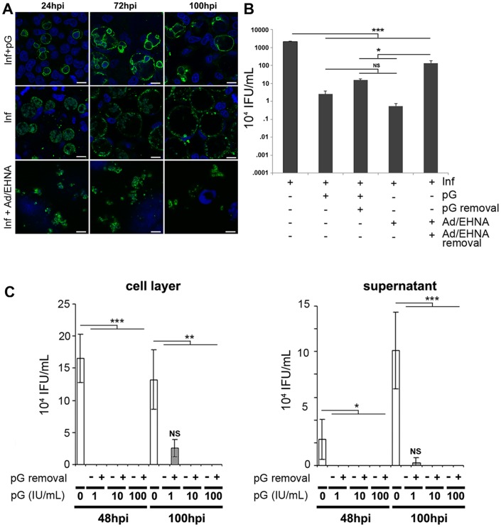
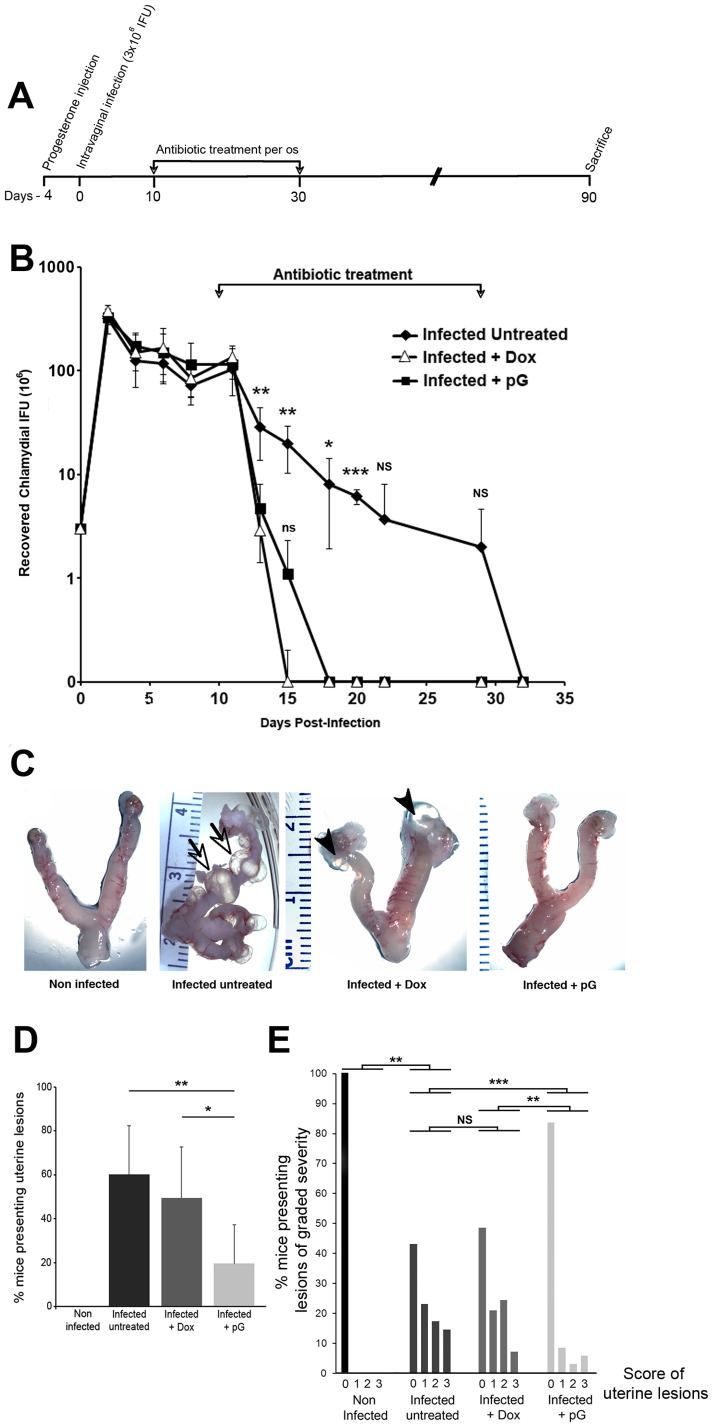
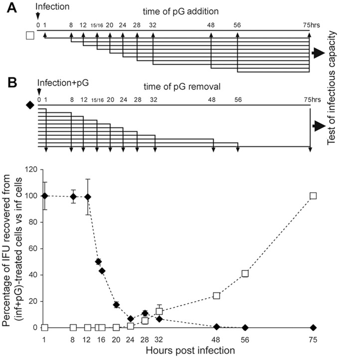
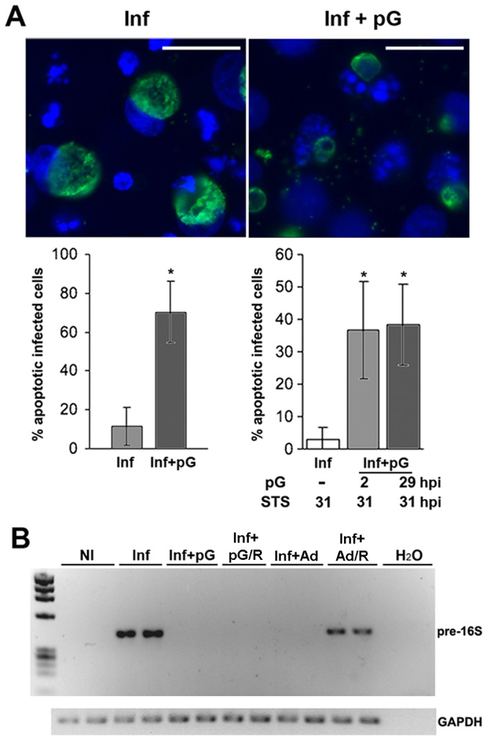
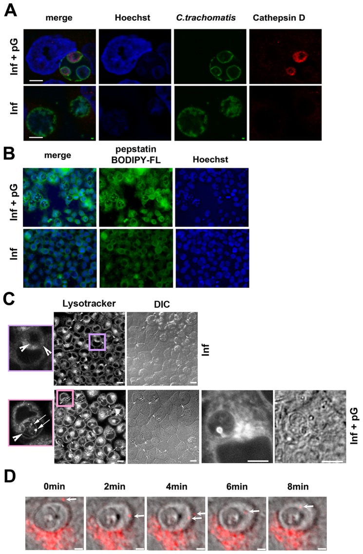
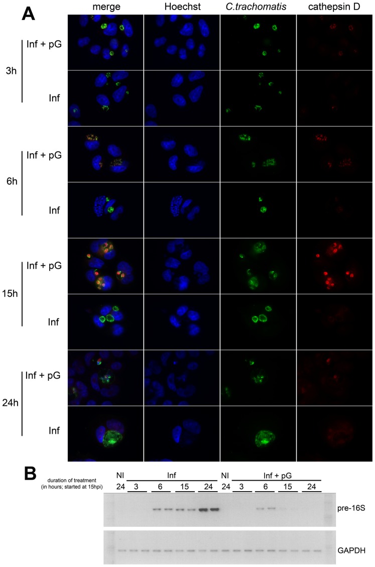
The obligate intracellular bacterium Chlamydia exists as two distinct forms. Elementary bodies (EBs) are infectious and extra-cellular, whereas reticulate bodies (RBs) replicate within a specialized intracellular compartment termed an 'inclusion'. Alternative persistent intra-cellular forms can be induced in culture by diverse stimuli such as IFNγ or adenosine/EHNA. They do not grow or divide but revive upon withdrawal of the stimulus and are implicated in several widespread human diseases through ill-defined in vivo mechanisms. β-Lactam antibiotics have also been claimed to induce persistence in vitro. The present report shows that upon penicillin G (pG) treatment, inclusions grow as fast as those in infected control cells. After removal of pG, Chlamydia do not revert to RBs. These effects are independent of host cell type, serovar, biovar and species of Chlamydia. Time-course experiments demonstrated that only RBs were susceptible to pG. pG-treated bacteria lost their control over host cell apoptotic pathways and no longer expressed pre-16S rRNA, in contrast to persistent bacteria induced with adenosine/EHNA. Confocal and live-video microscopy showed that bacteria within the inclusion fused with lysosomal compartments in pG-treated cells. That leads to recruitment of cathepsin D as early as 3 h post pG treatment, an event preceding bacterial death by several hours. These data demonstrate that pG treatment of cultured cells infected with Chlamydia results in the degradation of the bacteria. In addition we show that pG is significantly more efficient than doxycycline at preventing genital inflammatory lesions in C. muridarum-C57Bl/6 infected mice. These in vivo results support the physiological relevance of our findings and their potential therapeutic applications.
专性胞内细菌衣原体以两种不同形式存在。原体(EBs)具有传染性且存在于细胞外,而网状体(RBs)在一个称为“包涵体”的特殊胞内区室中进行复制。通过多种刺激因素,如γ干扰素或腺苷/依他尼酸(EHNA),可在培养物中诱导出替代性的持续性胞内形式。它们不生长也不分裂,但在刺激因素撤除后会复苏,并通过尚不明确的体内机制与多种广泛流行的人类疾病有关。β-内酰胺类抗生素也被认为可在体外诱导持续性。本报告显示,用青霉素G(pG)处理后,包涵体的生长速度与感染对照细胞中的包涵体一样快。去除pG后,衣原体不会恢复为网状体。这些效应与宿主细胞类型、血清型、生物变种以及衣原体的种类无关。时间进程实验表明,只有网状体对pG敏感。与用腺苷/EHNA诱导的持续性细菌相比,经pG处理的细菌失去了对宿主细胞凋亡途径的控制,且不再表达16S前体rRNA。共聚焦显微镜和实时视频显微镜显示,经pG处理的细胞中,包涵体内的细菌与溶酶体区室融合。这导致早在pG处理后3小时就募集组织蛋白酶D,这一事件比细菌死亡早几个小时。这些数据表明,用pG处理感染衣原体的培养细胞会导致细菌降解。此外,我们还表明,在预防感染鼠肺炎衣原体-C57Bl/6的小鼠发生生殖器炎性病变方面,pG比强力霉素显著更有效。这些体内实验结果支持了我们研究结果的生理相关性及其潜在的治疗应用价值。