Wang Defeng, Shi Lin, Liu Shangping, Hui Steve C N, Wang Yongjun, Cheng Jack C Y, Chu Winnie C W
Department of Imaging and Interventional Radiology, The Chinese University of Hong Kong, Shatin, New Territories, Hong Kong, China ; Research Center for Medical Image Computing, The Chinese University of Hong Kong, Shatin, New Territories, Hong Kong, China ; Shenzhen Research Institute, The Chinese University of Hong Kong, Shenzhen, China ; Department of Biomedical Engineering and Shun Hing Institute of Advanced Engineering, The Chinese University of Hong Kong, Shatin, New Territories, Hong Kong, China.
Department of Imaging and Interventional Radiology, The Chinese University of Hong Kong, Shatin, New Territories, Hong Kong, China ; Research Center for Medical Image Computing, The Chinese University of Hong Kong, Shatin, New Territories, Hong Kong, China.
PLoS One. 2013 Dec 20;8(12):e83767. doi: 10.1371/journal.pone.0083767. eCollection 2013.
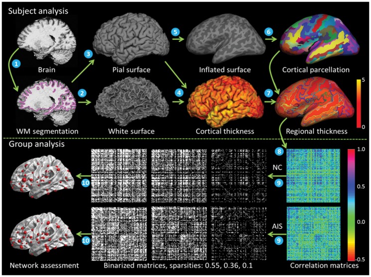
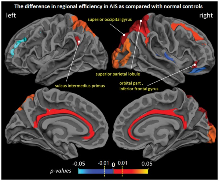
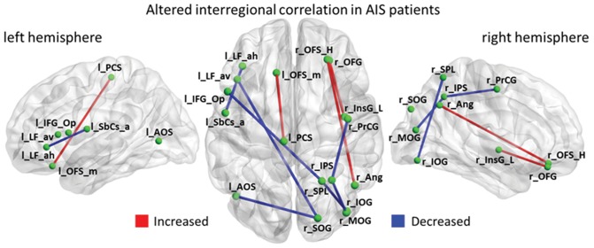
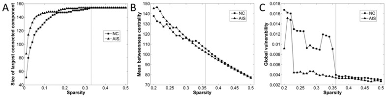
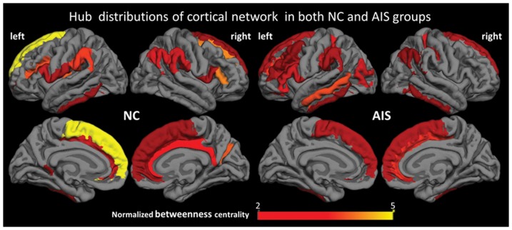
Adolescent idiopathic scoliosis (AIS) is a multifactorial disease affecting approximately 1-4% of teenagers especially girls at the age of 10-16, but its etiopathogenesis remains uncertain. Previous study has revealed that the cortical thickness in AIS patients is different from that in normal controls. Cortical thickness measurements are known to be strongly correlated between regions that are axonally connected. Hence, a hypothesis is proposed to study the possibility to demonstrate abnormal structural network revealed by cortical thickness in AIS patients. The aim of the study is to investigate abnormalities in the organization of the brain cortical network in AIS patients. This study included 42 girls with severe idiopathic scoliosis (14.7±1.3 years old) and 41 age-matched normal controls (NC, 14.6±1.4 years old). The brain cortex was partitioned into 154 cortical regions based on gyral and sulcal structure. The interregional connectivity was measured as the statistical correlations between the regional mean thicknesses across the subjects. We employed the graph theoretic analysis to examine the alteration in interregional correlation, small-world efficiency, hub distribution, and regional nodal characteristics in AIS patients. We demonstrated that the cortical network of AIS patients fully preserved the small-world architecture and organization, and further verified the hemispheric asymmetry of AIS brain. Our results indicated increased central role of temporal and occipital cortex and decreased central role of limbic cortex in AIS patients compared with controls. Furthermore, decreased structural connectivity between hemispheres and increased connectivity in several cortical regions were observed. The findings of the study reveal the pattern of structural network alteration in AIS brain, and would help in understanding the mechanism and etiopathogenesis of AIS.
青少年特发性脊柱侧凸(AIS)是一种多因素疾病,影响着约1%-4%的青少年,尤其是10至16岁的女孩,但其发病机制仍不确定。先前的研究表明,AIS患者的皮质厚度与正常对照组不同。已知轴突相连区域之间的皮质厚度测量值具有很强的相关性。因此,提出了一个假设,以研究通过AIS患者皮质厚度揭示异常结构网络的可能性。本研究的目的是调查AIS患者脑皮质网络组织的异常情况。本研究纳入了42名重度特发性脊柱侧凸女孩(14.7±1.3岁)和41名年龄匹配的正常对照(NC,14.6±1.4岁)。根据脑回和脑沟结构,将大脑皮质划分为154个皮质区域。区域间连通性通过受试者间区域平均厚度的统计相关性来衡量。我们采用图论分析来检查AIS患者区域间相关性、小世界效率、枢纽分布和区域节点特征的变化。我们证明,AIS患者的皮质网络完全保留了小世界架构和组织,并进一步验证了AIS大脑的半球不对称性。我们的结果表明,与对照组相比,AIS患者颞叶和枕叶皮质的中心作用增强,边缘皮质的中心作用减弱。此外,还观察到半球间结构连通性降低,以及几个皮质区域的连通性增加。该研究结果揭示了AIS大脑结构网络改变的模式,将有助于理解AIS的机制和发病机制。