• 文献检索
  • 文档翻译
  • 深度研究
  • 学术资讯
  • Suppr Zotero 插件Zotero 插件
  • 邀请有礼
  • 套餐&价格
  • 历史记录
应用&插件
Suppr Zotero 插件Zotero 插件浏览器插件Mac 客户端Windows 客户端微信小程序
定价
高级版会员购买积分包购买API积分包
服务
文献检索文档翻译深度研究API 文档MCP 服务
关于我们
关于 Suppr公司介绍联系我们用户协议隐私条款
关注我们

Suppr 超能文献

核心技术专利:CN118964589B侵权必究
粤ICP备2023148730 号-1Suppr @ 2026

文献检索

告别复杂PubMed语法,用中文像聊天一样搜索,搜遍4000万医学文献。AI智能推荐,让科研检索更轻松。

立即免费搜索

文件翻译

保留排版,准确专业,支持PDF/Word/PPT等文件格式,支持 12+语言互译。

免费翻译文档

深度研究

AI帮你快速写综述,25分钟生成高质量综述,智能提取关键信息,辅助科研写作。

立即免费体验

使用256层计算机断层扫描对三磷酸腺苷负荷全心动态心肌灌注成像进行定性和定量评估。

Qualitative and quantitative assessment of adenosine triphosphate stress whole-heart dynamic myocardial perfusion imaging using 256-slice computed tomography.

作者信息

Kurata Akira, Kawaguchi Naoto, Kido Teruhito, Inoue Katsuji, Suzuki Jun, Ogimoto Akiyoshi, Funada Jun-ichi, Higaki Jitsuo, Miyagawa Masao, Vembar Mani, Mochizuki Teruhito

机构信息

Department of Diagnostic and Therapeutic Radiology, Ehime University Graduate School of Medicine, Ehime, Japan.

Department of Integrated Medicine and Informatics, Ehime University Graduate School of Medicine, Ehime, Japan.

出版信息

PLoS One. 2013 Dec 23;8(12):e83950. doi: 10.1371/journal.pone.0083950. eCollection 2013.

DOI:10.1371/journal.pone.0083950
PMID:24376774
原文链接:https://pmc.ncbi.nlm.nih.gov/articles/PMC3871670/
Abstract

BACKGROUND

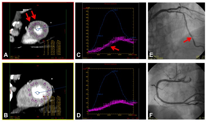
The aim of this study was to investigate the correlation of the qualitative transmural extent of hypoperfusion areas (HPA) using stress dynamic whole-heart computed tomography perfusion (CTP) imaging by 256-slice CT with CTP-derived myocardial blood flow (MBF) for the estimation of the severity of coronary artery stenosis.

METHODS AND RESULTS

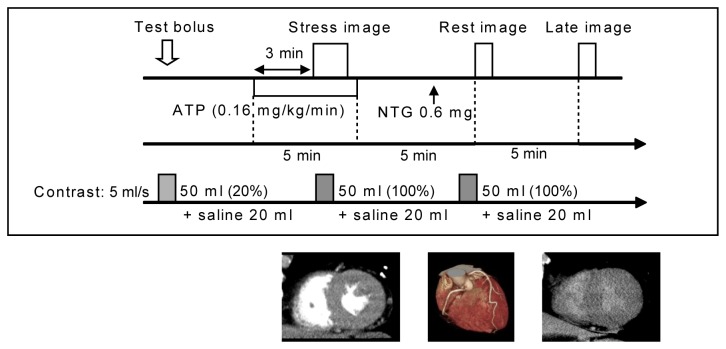
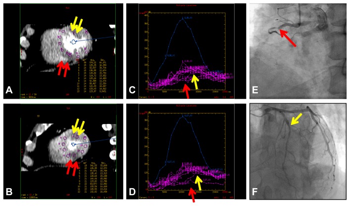
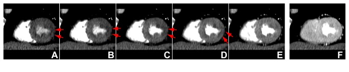
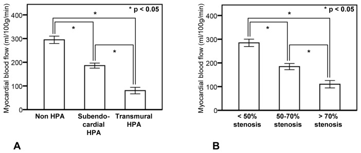
Eleven patients underwent adenosine triphosphate (0.16 mg/kg/min, 5 min) stress dynamic CTP by 256-slice CT (coverage: 8 cm, 0.27 s/rotation), and 9 of the 11 patients underwent coronary angiography (CAG). Stress dynamic CTP (whole-heart datasets over 30 consecutive heart beats in systole without spatial and temporal gaps) was acquired with prospective ECG gating (effective radiation dose: 10.4 mSv). The extent of HPAs was visually graded using a 3-point score (normal, subendocardial, transmural). MBF (ml/100g/min) was measured by deconvolution. Differences in MBF (mean ± standard error) according to HPA and CAG results were evaluated. In 27 regions (3 major coronary territories in 9 patients), 11 coronary stenoses (> 50% reduction in diameter) were observed. In 353 myocardial segments, HPA was significantly related to MBF (P < 0.05; normal 295 ± 94; subendocardial 186 ± 67; and transmural 80 ± 53). Coronary territory analysis revealed a significant relationship between coronary stenosis severity and MBF (P < 0.05; non-significant stenosis [< 50%], 284 ± 97; moderate stenosis [50-70%], 184 ± 74; and severe stenosis [> 70%], 119 ± 69).

CONCLUSION

The qualitative transmural extent of HPA using stress whole-heart dynamic CTP imaging by 256-slice CT exhibits a good correlation with quantitative CTP-derived MBF and may aid in assessing the hemodynamic significance of coronary artery disease.

摘要

背景

本研究旨在通过256层CT的应力动态全心脏计算机断层扫描灌注(CTP)成像,研究灌注减低区(HPA)的定性透壁范围与CTP衍生的心肌血流量(MBF)之间的相关性,以评估冠状动脉狭窄的严重程度。

方法与结果

11例患者接受了256层CT的三磷酸腺苷(0.16mg/kg/min,5分钟)应力动态CTP检查(覆盖范围:8cm,0.27秒/旋转),11例患者中的9例接受了冠状动脉造影(CAG)。采用前瞻性心电图门控获取应力动态CTP(收缩期连续30个心跳的全心脏数据集,无空间和时间间隙)(有效辐射剂量:10.4mSv)。使用3分评分(正常、心内膜下、透壁)对HPA的范围进行视觉分级。通过去卷积测量MBF(ml/100g/min)。评估根据HPA和CAG结果的MBF差异(平均值±标准误差)。在27个区域(9例患者的3个主要冠状动脉区域),观察到11处冠状动脉狭窄(直径减少>50%)。在353个心肌节段中,HPA与MBF显著相关(P<0.05;正常295±94;心内膜下186±67;透壁80±53)。冠状动脉区域分析显示冠状动脉狭窄严重程度与MBF之间存在显著关系(P<0.05;非显著狭窄[<50%],284±97;中度狭窄[50-70%],184±74;重度狭窄[>70%],119±69)。

结论

使用256层CT的应力全心脏动态CTP成像对HPA进行定性透壁范围分析,与定量CTP衍生的MBF具有良好的相关性,可能有助于评估冠状动脉疾病的血流动力学意义。

https://cdn.ncbi.nlm.nih.gov/pmc/blobs/3199/3871670/d50bfe59f07a/pone.0083950.g005.jpg
https://cdn.ncbi.nlm.nih.gov/pmc/blobs/3199/3871670/e0ba26b3f9b0/pone.0083950.g001.jpg
https://cdn.ncbi.nlm.nih.gov/pmc/blobs/3199/3871670/743ed1bef716/pone.0083950.g002.jpg
https://cdn.ncbi.nlm.nih.gov/pmc/blobs/3199/3871670/e2ad5e734b95/pone.0083950.g003.jpg
https://cdn.ncbi.nlm.nih.gov/pmc/blobs/3199/3871670/a881b68739cd/pone.0083950.g004.jpg
https://cdn.ncbi.nlm.nih.gov/pmc/blobs/3199/3871670/d50bfe59f07a/pone.0083950.g005.jpg

相似文献

1
Qualitative and quantitative assessment of adenosine triphosphate stress whole-heart dynamic myocardial perfusion imaging using 256-slice computed tomography.使用256层计算机断层扫描对三磷酸腺苷负荷全心动态心肌灌注成像进行定性和定量评估。
PLoS One. 2013 Dec 23;8(12):e83950. doi: 10.1371/journal.pone.0083950. eCollection 2013.
2
Comprehensive morphologic and functional imaging of heart transplant patients: first experience with dynamic perfusion CT.心脏移植患者的全面形态学和功能成像:动态灌注 CT 的初步经验。
Eur Radiol. 2018 Oct;28(10):4111-4121. doi: 10.1007/s00330-018-5436-9. Epub 2018 Apr 30.
3
Diagnostic value of transmural perfusion ratio derived from dynamic CT-based myocardial perfusion imaging for the detection of haemodynamically relevant coronary artery stenosis.基于动态CT心肌灌注成像的透壁灌注率对检测血流动力学相关冠状动脉狭窄的诊断价值。
Eur Radiol. 2017 Jun;27(6):2309-2316. doi: 10.1007/s00330-016-4567-0. Epub 2016 Oct 4.
4
Feasibility of dynamic myocardial CT perfusion using single-source 64-row CT.应用单源 64 排 CT 进行动态心肌 CT 灌注的可行性研究。
J Cardiovasc Comput Tomogr. 2019 Jan-Feb;13(1):55-61. doi: 10.1016/j.jcct.2018.10.003. Epub 2018 Oct 6.
5
Coronary artery stenosis-related perfusion ratio using dynamic computed tomography myocardial perfusion imaging: a pilot for identification of hemodynamically significant coronary artery disease.采用动态 CT 心肌灌注成像评估冠状动脉狭窄相关的灌注比值:识别有血流动力学意义的冠心病的初步研究。
Cardiovasc Interv Ther. 2020 Oct;35(4):327-335. doi: 10.1007/s12928-019-00627-4. Epub 2019 Oct 19.
6
Accuracy of dynamic computed tomography adenosine stress myocardial perfusion imaging in estimating myocardial blood flow at various degrees of coronary artery stenosis using a porcine animal model.应用猪动物模型评估动态 CT 腺苷负荷心肌灌注成像在不同程度冠状动脉狭窄时估算心肌血流的准确性。
Invest Radiol. 2012 Jan;47(1):71-7. doi: 10.1097/RLI.0b013e31823fd42b.
7
Computed tomography myocardial perfusion vs O-water positron emission tomography and fractional flow reserve.计算机断层扫描心肌灌注与O-水正电子发射断层扫描及血流储备分数的比较
Eur Radiol. 2017 Mar;27(3):1114-1124. doi: 10.1007/s00330-016-4404-5. Epub 2016 Jun 22.
8
Classification of hemodynamically significant stenoses from dynamic CT perfusion and CTA myocardial territories.基于动态CT灌注和CTA心肌区域对血流动力学显著狭窄进行分类。
Med Phys. 2017 Apr;44(4):1347-1358. doi: 10.1002/mp.12126. Epub 2017 Mar 22.
9
Stress and rest dynamic myocardial perfusion imaging by evaluation of complete time-attenuation curves with dual-source CT.应用双源 CT 评估完整的时间衰减曲线进行应激和静息心肌灌注成像。
JACC Cardiovasc Imaging. 2010 Aug;3(8):811-20. doi: 10.1016/j.jcmg.2010.05.009.
10
Incremental Prognostic Value of Myocardial Blood Flow Quantified With Stress Dynamic Computed Tomography Perfusion Imaging.压力动态计算机断层扫描灌注成像定量心肌血流的增量预后价值。
JACC Cardiovasc Imaging. 2019 Jul;12(7 Pt 2):1379-1387. doi: 10.1016/j.jcmg.2018.05.021. Epub 2018 Jul 18.

引用本文的文献

1
Dynamic CT Myocardial Perfusion: The Role of Functional Evaluation in the Diagnosis of Coronary Artery Disease.动态CT心肌灌注:功能评估在冠状动脉疾病诊断中的作用
J Clin Med. 2023 Nov 13;12(22):7062. doi: 10.3390/jcm12227062.
2
A Novel Quantitative Parameter for Static Myocardial Computed Tomography: Myocardial Perfusion Ratio to the Aorta.一种用于静态心肌计算机断层扫描的新型定量参数:心肌灌注与主动脉比值。
J Clin Med. 2022 Mar 25;11(7):1816. doi: 10.3390/jcm11071816.
3
Role of DECT in coronary artery disease: a comparative study with ICA and SPECT.

本文引用的文献

1
Quantification of myocardial blood flow by adenosine-stress CT perfusion imaging in pigs during various degrees of stenosis correlates well with coronary artery blood flow and fractional flow reserve.腺苷负荷 CT 灌注成像定量检测不同狭窄程度猪心肌血流量与冠状动脉血流和血流储备分数相关性良好。
Eur Heart J Cardiovasc Imaging. 2013 Apr;14(4):331-8. doi: 10.1093/ehjci/jes150. Epub 2012 Jul 26.
2
Cardiovascular events in Japan. Lessons from the J-ACCESS multicenter prognostic study using myocardial perfusion imaging.日本的心血管事件。使用心肌灌注成像的 J-ACCESS 多中心预后研究的经验教训。
Circ J. 2012;76(6):1313-21. doi: 10.1253/circj.cj-12-0260. Epub 2012 Apr 26.
3
DECT 在冠心病中的作用:与 ICA 和 SPECT 的对比研究。
Diagn Interv Radiol. 2020 Sep;26(5):420-428. doi: 10.5152/dir.2020.18569.
4
A noteworthy clue for diagnosing non-ST-elevation myocardial infarction by computed tomography without ECG synchronization: a case report.值得注意的通过非心电图同步计算机断层扫描诊断非 ST 段抬高型心肌梗死的线索:病例报告。
BMC Cardiovasc Disord. 2020 May 25;20(1):244. doi: 10.1186/s12872-020-01512-2.
5
Fusion of Three-Dimensional Echocardiographic Regional Myocardial Strain with Cardiac Computed Tomography for Noninvasive Evaluation of the Hemodynamic Impact of Coronary Stenosis in Patients with Chest Pain.三维超声心动图局部心肌应变与心脏计算机断层扫描融合技术用于评估胸痛患者冠状动脉狭窄的血液动力学影响的非侵入性评价。
J Am Soc Echocardiogr. 2018 Jun;31(6):664-673. doi: 10.1016/j.echo.2018.01.019. Epub 2018 Mar 22.
6
Evaluation of an adaptive detector collimation for prospectively ECG-triggered coronary CT angiography with third-generation dual-source CT.评价第三代双源 CT 前瞻性心电门控冠状动脉 CT 血管造影自适应探测器准直的应用。
Eur Radiol. 2018 May;28(5):2143-2150. doi: 10.1007/s00330-017-5177-1. Epub 2017 Dec 7.
7
Comprehensive Assessment of Coronary Artery Disease by Using First-Pass Analysis Dynamic CT Perfusion: Validation in a Swine Model.利用首过分析动态CT灌注对冠状动脉疾病进行综合评估:在猪模型中的验证
Radiology. 2018 Jan;286(1):93-102. doi: 10.1148/radiol.2017162821. Epub 2017 Oct 23.
8
Impact of knowledge-based iterative model reconstruction on myocardial late iodine enhancement in computed tomography and comparison with cardiac magnetic resonance.基于知识的迭代模型重建对计算机断层扫描中心肌晚期碘增强的影响及与心脏磁共振成像的比较
Int J Cardiovasc Imaging. 2017 Oct;33(10):1609-1618. doi: 10.1007/s10554-017-1137-8. Epub 2017 Apr 13.
9
Functional Assessment of Coronary Artery Disease Using Whole-Heart Dynamic Computed Tomographic Perfusion.使用全心动态计算机断层扫描灌注技术对冠状动脉疾病进行功能评估。
Circ Cardiovasc Imaging. 2016 Dec;9(12). doi: 10.1161/CIRCIMAGING.116.005325.
10
Differentiation of myocardial ischemia and infarction assessed by dynamic computed tomography perfusion imaging and comparison with cardiac magnetic resonance and single-photon emission computed tomography.动态计算机断层灌注成像评估心肌缺血和梗死的鉴别诊断,并与心脏磁共振和单光子发射计算机断层扫描比较。
Eur Radiol. 2016 Nov;26(11):3790-3801. doi: 10.1007/s00330-016-4238-1. Epub 2016 Feb 6.
Adenosine-stress dynamic myocardial perfusion imaging with second-generation dual-source CT: comparison with conventional catheter coronary angiography and SPECT nuclear myocardial perfusion imaging.
腺苷负荷动态心肌灌注成像双源 CT 第二代:与常规导管冠状动脉造影和 SPECT 核素心肌灌注成像比较。
AJR Am J Roentgenol. 2012 Mar;198(3):521-9. doi: 10.2214/AJR.11.7830.
4
Assessment of cardiac computed tomography-myocardial perfusion imaging - promise and challenges - .心脏计算机断层扫描心肌灌注成像评估——前景与挑战——。
Circ J. 2012;76(3):544-52. doi: 10.1253/circj.cj-11-1427. Epub 2012 Feb 11.
5
Quantitative myocardial perfusion analysis using multi-row detector CT in acute myocardial infarction.采用多排探测器 CT 对急性心肌梗死进行定量心肌灌注分析。
Heart. 2012 Apr;98(7):566-72. doi: 10.1136/heartjnl-2011-300915. Epub 2012 Jan 27.
6
Rapid detection of coronary artery stenoses with real-time perfusion echocardiography during regadenoson stress.实时灌注超声心动图在瑞加德松应激时快速检测冠状动脉狭窄。
Circ Cardiovasc Imaging. 2011 Nov;4(6):628-35. doi: 10.1161/CIRCIMAGING.111.966341. Epub 2011 Sep 23.
7
Non-invasive assessment of functionally relevant coronary artery stenoses with quantitative CT perfusion: preliminary clinical experiences.采用定量 CT 灌注技术无创性评估有功能意义的冠状动脉狭窄:初步临床经验。
Eur Radiol. 2012 Jan;22(1):39-50. doi: 10.1007/s00330-011-2260-x. Epub 2011 Sep 21.
8
Combination of a low-tube-voltage technique with hybrid iterative reconstruction (iDose) algorithm at coronary computed tomographic angiography.在冠状动脉计算机断层血管造影中低管电压技术与混合迭代重建(iDose)算法的联合应用。
J Comput Assist Tomogr. 2011 Jul-Aug;35(4):480-5. doi: 10.1097/RCT.0b013e31821fee94.
9
Transmural perfusion gradient in adenosine triphosphate stress myocardial perfusion computed tomography.腺苷三磷酸负荷心肌灌注 CT 中的跨壁灌注梯度。
Circ J. 2011;75(8):1905-12. doi: 10.1253/circj.cj-10-1144. Epub 2011 Jun 21.
10
Stress and rest dynamic myocardial perfusion imaging by evaluation of complete time-attenuation curves with dual-source CT.应用双源 CT 评估完整的时间衰减曲线进行应激和静息心肌灌注成像。
JACC Cardiovasc Imaging. 2010 Aug;3(8):811-20. doi: 10.1016/j.jcmg.2010.05.009.