Heidari Mohammad Hassan, Amini Abdollah, Bahrami Zohreh, Shahriari Ali, Movafag Abolfazle, Heidari Reihane
Department of Biology and Anatomical Sciences, Faculty of Medicine, Shahid Beheshti University of Medical Sciences, Tehran, Iran.
Department of Anesthesiology, Roozbeh Hospital, Tehran University of Medical Sciences, P.O. Box 1417653761, Tehran, Iran.
Neurol Res Int. 2013;2013:290414. doi: 10.1155/2013/290414. Epub 2013 Nov 28.
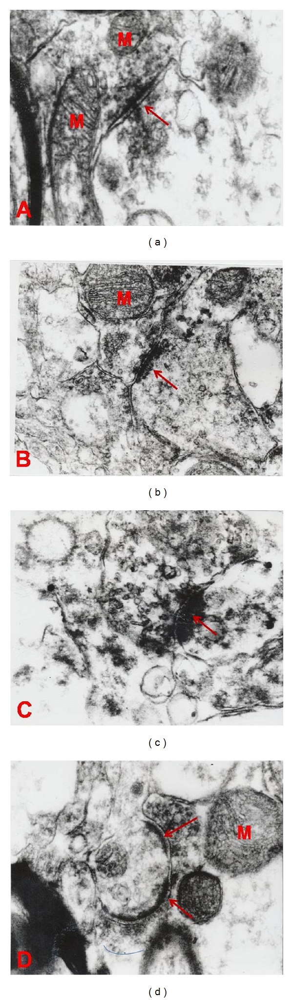
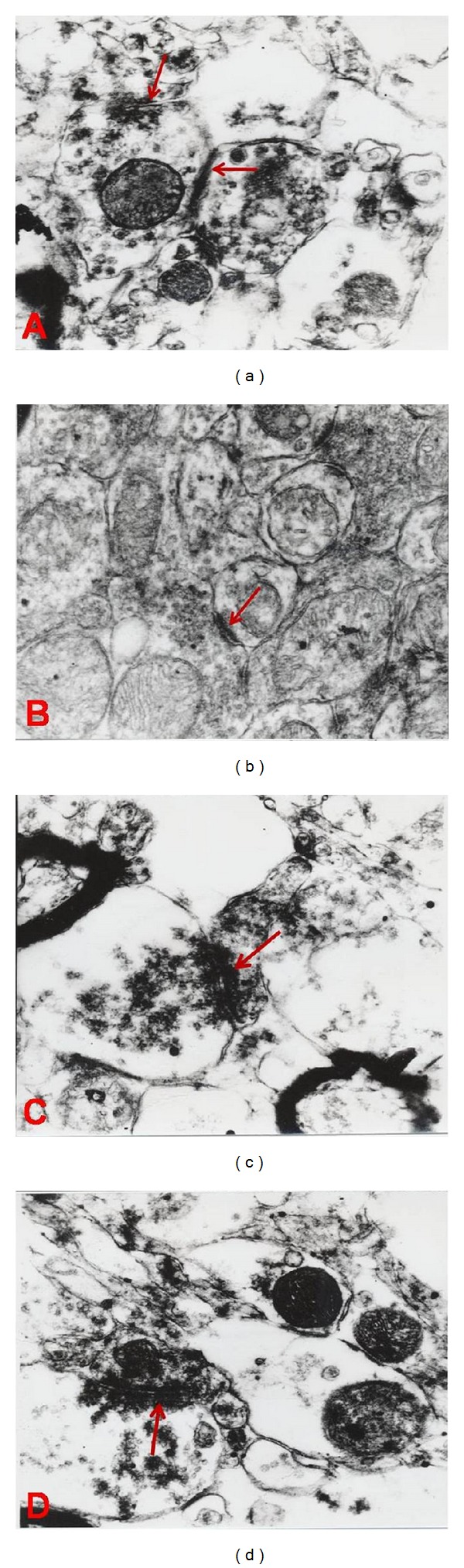
It is well known that the synapses undergo some changes in the brain during the course of normal life and under certain pathological or experimental circumstances. One of the main goals of numerous researchers has been to find the reasons for these structural changes. In the present study, we investigated the effects of chronic morphine consumption on synaptic plasticity, postsynaptic density thickness, and synaptic curvatures of hippocampus CA1 area of rats. So for reaching these goals, 24 N-Mary male rats were randomly divided into three groups, morphine (n = 8), placebo (n = 8), and control (n = 8) groups. In the morphine group, complex of morphine (0.1, 0.2, 0.3, and 0.4) mg/mL and in the placebo (sucrose) group complex of sucrose (% 0.3) were used for 21 days. After the end of drug treatment the animals were scarified and perfused intracardinally and finally the CA1 hippocampal samples were taken for ultrastructural studies, and then the obtained data were analyzed by SPSS and one-way analysis of variance. Our data indicated that synaptic numbers per nm(3) change significantly in morphine group compared to the other two groups (placebo and control) (P < 0.001) and also statistical analysis revealed a significant difference between groups in terms of thickness of postsynaptic density (P < 0.001) and synaptic curvature (P < 0.007). It seems that morphine dependence in rats plays a main role in the ultrastructural changes of hippocampus.
众所周知,在正常生活过程中以及在某些病理或实验情况下,大脑中的突触会发生一些变化。众多研究人员的主要目标之一就是找出这些结构变化的原因。在本研究中,我们调查了长期服用吗啡对大鼠海马CA1区突触可塑性、突触后致密物厚度和突触曲率的影响。因此,为了实现这些目标,将24只雄性N-Mary大鼠随机分为三组,即吗啡组(n = 8)、安慰剂组(n = 8)和对照组(n = 8)。在吗啡组中,使用吗啡(0.1、0.2、0.3和0.4)mg/mL的复合物,在安慰剂(蔗糖)组中使用蔗糖(0.3%)的复合物,持续21天。药物治疗结束后,对动物实施安乐死并进行心脏灌注,最后取海马CA1区样本进行超微结构研究,然后用SPSS软件和单因素方差分析对所得数据进行分析。我们的数据表明,与其他两组(安慰剂组和对照组)相比,吗啡组每立方纳米的突触数量有显著变化(P < 0.001),而且统计分析显示,在突触后致密物厚度(P < 0.001)和突触曲率(P < 0.007)方面,各组之间存在显著差异。似乎大鼠对吗啡的依赖在海马的超微结构变化中起主要作用。