Eckhard A, Müller M, Salt A, Smolders J, Rask-Andersen H, Löwenheim H
Hearing Research Center, Department of Otorhinolaryngology-Head & Neck Surgery, University of Tübingen Medical Centre, Elfriede-Aulhorn-Strasse 5, 72076, Tübingen, Germany.
Pflugers Arch. 2014 Oct;466(10):1963-85. doi: 10.1007/s00424-013-1421-y. Epub 2014 Jan 3.
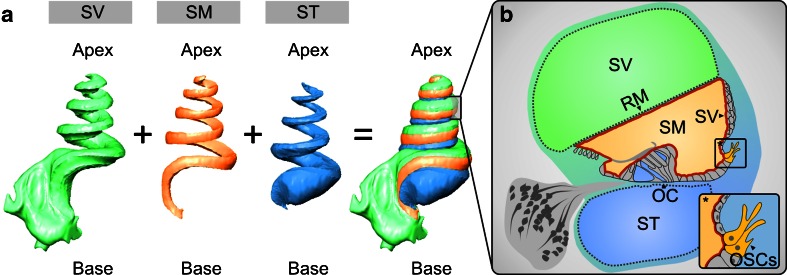
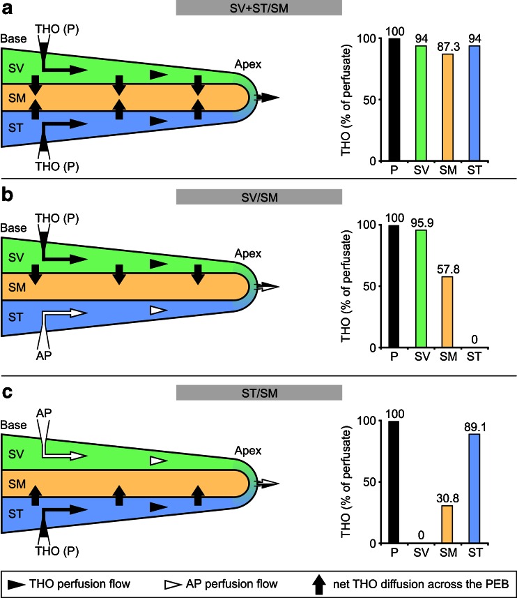
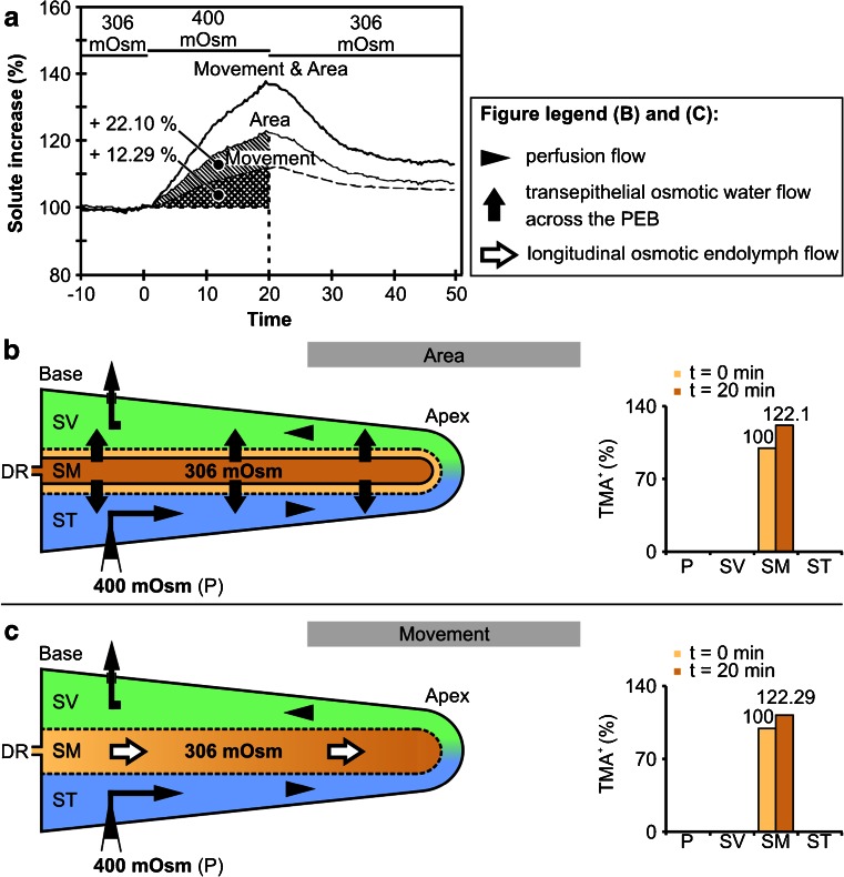
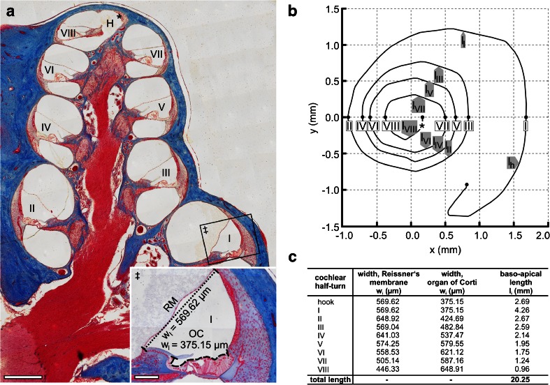
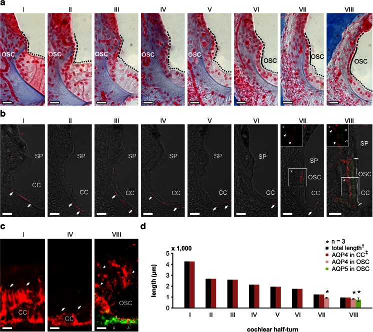
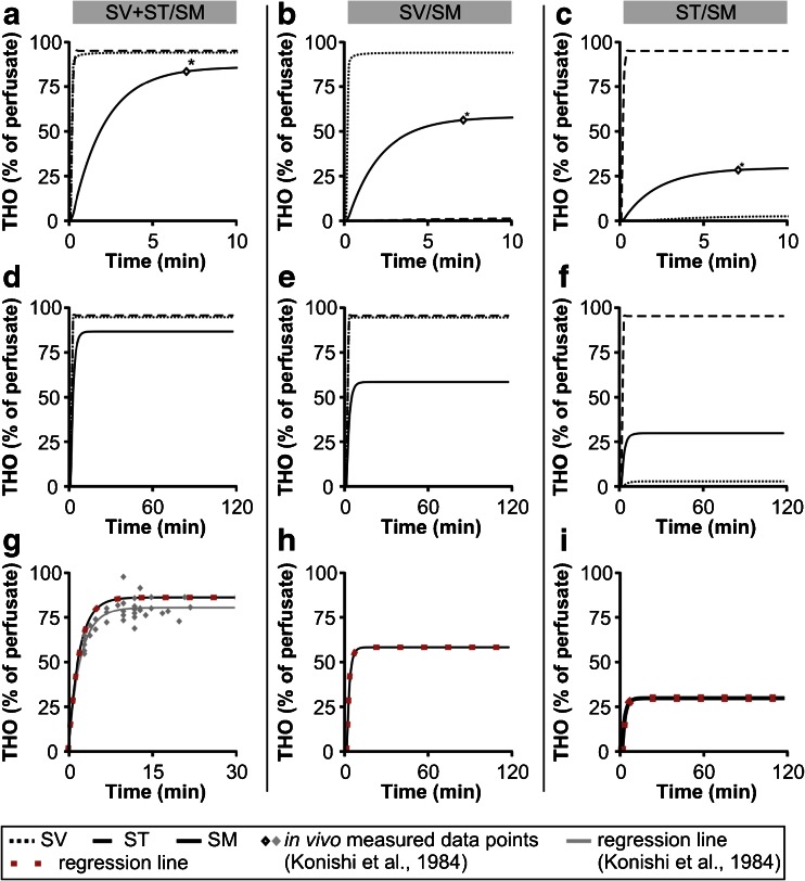
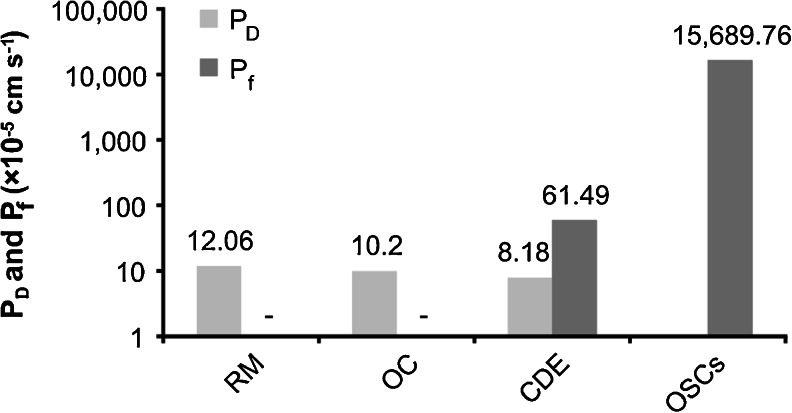
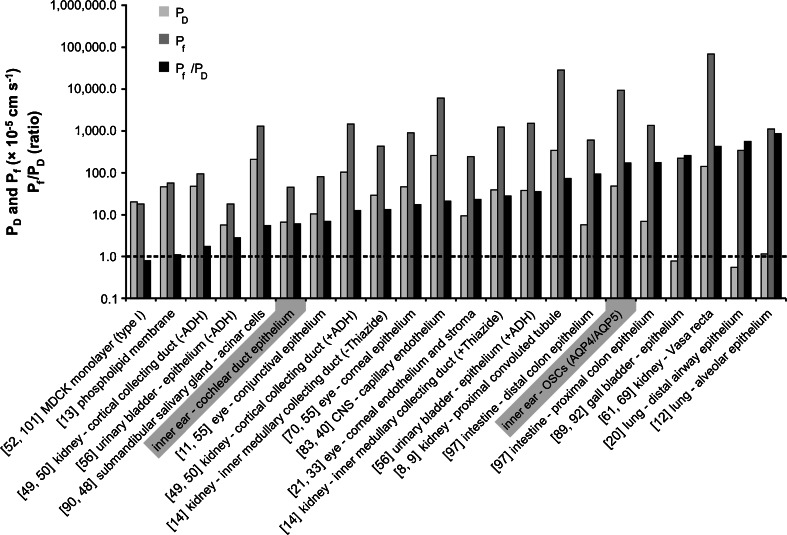
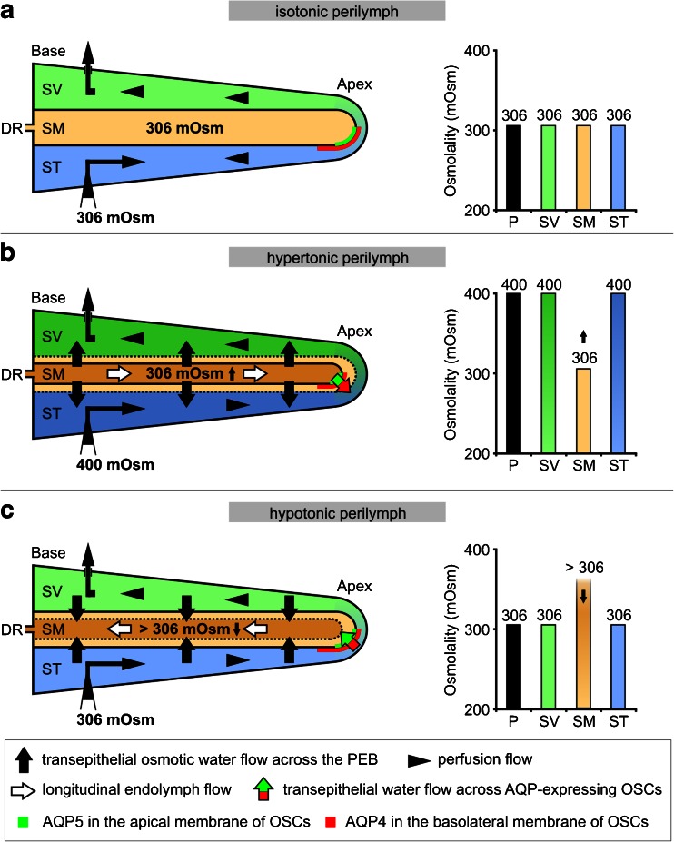
The cochlear duct epithelium (CDE) constitutes a tight barrier that effectively separates the inner ear fluids, endolymph and perilymph, thereby maintaining distinct ionic and osmotic gradients that are essential for auditory function. However, in vivo experiments have demonstrated that the CDE allows for rapid water exchange between fluid compartments. The molecular mechanism governing water permeation across the CDE remains elusive. We computationally determined the diffusional (PD) and osmotic (Pf) water permeability coefficients for the mammalian CDE based on in silico simulations of cochlear water dynamics integrating previously derived in vivo experimental data on fluid flow with expression sites of molecular water channels (aquaporins, AQPs). The PD of the entire CDE (PD = 8.18 × 10(-5) cm s(-1)) and its individual partitions including Reissner's membrane (PD = 12.06 × 10(-5) cm s(-1)) and the organ of Corti (PD = 10.2 × 10(-5) cm s(-1)) were similar to other epithelia with AQP-facilitated water permeation. The Pf of the CDE (Pf = 6.15 × 10(-4) cm s(-1)) was also in the range of other epithelia while an exceptionally high Pf was determined for an epithelial subdomain of outer sulcus cells in the cochlear apex co-expressing AQP4 and AQP5 (OSCs; Pf = 156.90 × 10(-3) cm s(-1)). The Pf/PD ratios of the CDE (Pf/PD = 7.52) and OSCs (Pf/PD = 242.02) indicate an aqueous pore-facilitated water exchange and reveal a high-transfer region or "water shunt" in the cochlear apex. This "water shunt" explains experimentally determined phenomena of endolymphatic longitudinal flow towards the cochlear apex. The water permeability coefficients of the CDE emphasise the physiological and pathophysiological relevance of water dynamics in the cochlea in particular for endolymphatic hydrops and Ménière's disease.
耳蜗管上皮(CDE)构成了一个紧密的屏障,有效地分隔了内耳的内淋巴和外淋巴液,从而维持了对听觉功能至关重要的独特离子和渗透压梯度。然而,体内实验表明,CDE允许液体隔室之间快速进行水交换。控制水透过CDE的分子机制仍然不清楚。我们基于耳蜗水动力学的计算机模拟,结合先前获得的关于流体流动的体内实验数据和分子水通道(水通道蛋白,AQPs)的表达位点,通过计算确定了哺乳动物CDE的扩散(PD)和渗透(Pf)水渗透系数。整个CDE的PD(PD = 8.18×10⁻⁵ cm s⁻¹)及其各个部分,包括Reissner膜(PD = 12.06×10⁻⁵ cm s⁻¹)和柯蒂氏器(PD = 10.2×10⁻⁵ cm s⁻¹),与其他有AQP促进水渗透的上皮组织相似。CDE的Pf(Pf = 6.15×10⁻⁴ cm s⁻¹)也在其他上皮组织的范围内,而在耳蜗顶端共表达AQP4和AQP5的外沟细胞上皮亚域(OSCs;Pf = 156.90×10⁻³ cm s⁻¹)测定出了异常高的Pf。CDE(Pf/PD = 7.52)和OSCs(Pf/PD = 242.02)的Pf/PD比值表明存在水孔促进的水交换,并揭示了耳蜗顶端的一个高转运区域或“水分流”。这种“水分流”解释了实验确定的内淋巴向耳蜗顶端纵向流动的现象。CDE的水渗透系数强调了耳蜗中水动力学在生理和病理生理方面的相关性,特别是对内淋巴积水和梅尼埃病而言。