Section of Otolaryngology-Head and Neck Surgery, University of Chicago, Chicago, Illinois, USA.
Department of ENT-Head and Neck Surgery, University Hospital RWTH Aachen, Aachen, Germany.
Otol Neurotol. 2020 Apr;41(4):e507-e515. doi: 10.1097/MAO.0000000000002535.
Outer sulcus cell features and distribution are hypothesized to differ throughout regions of the human cochlea and between diseased and normal specimens.
Outer sulcus cells play a role in inner ear fluid homeostasis. However, their anatomy and distribution in the human are not well described.
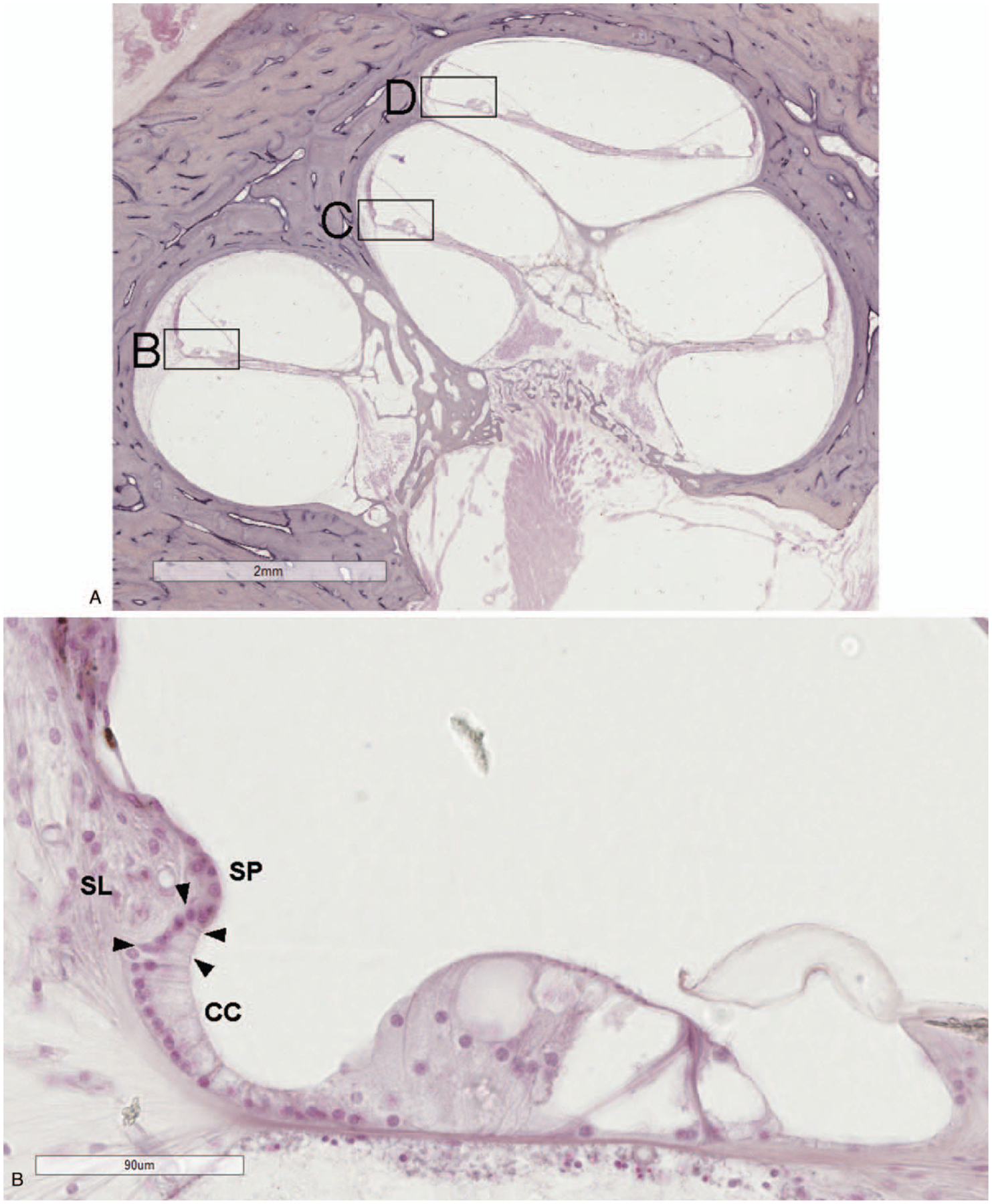
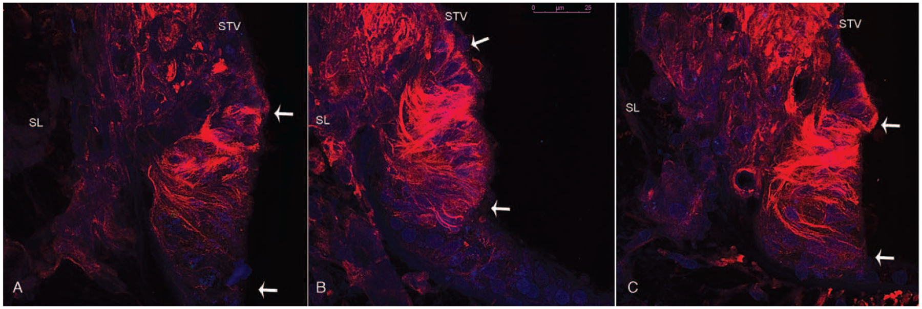
Temporal bone specimens with normal hearing (n = 10), Menière's disease (n = 10), presbycusis with flat audiograms (n = 4), and presbycusis with sloping audiograms (n = 5) were examined by light microscopy. Outer sulcus cells were assessed quantitatively and qualitatively in each cochlear turn. One specimen was stained for tubulin immunofluorescence and imaged using confocal microscopy.
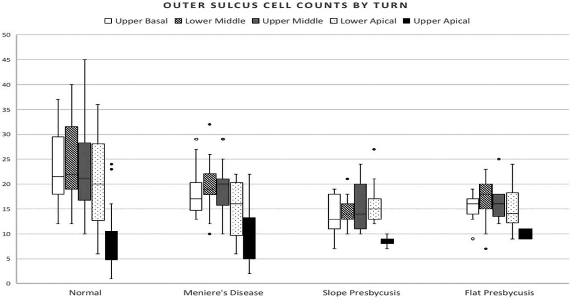
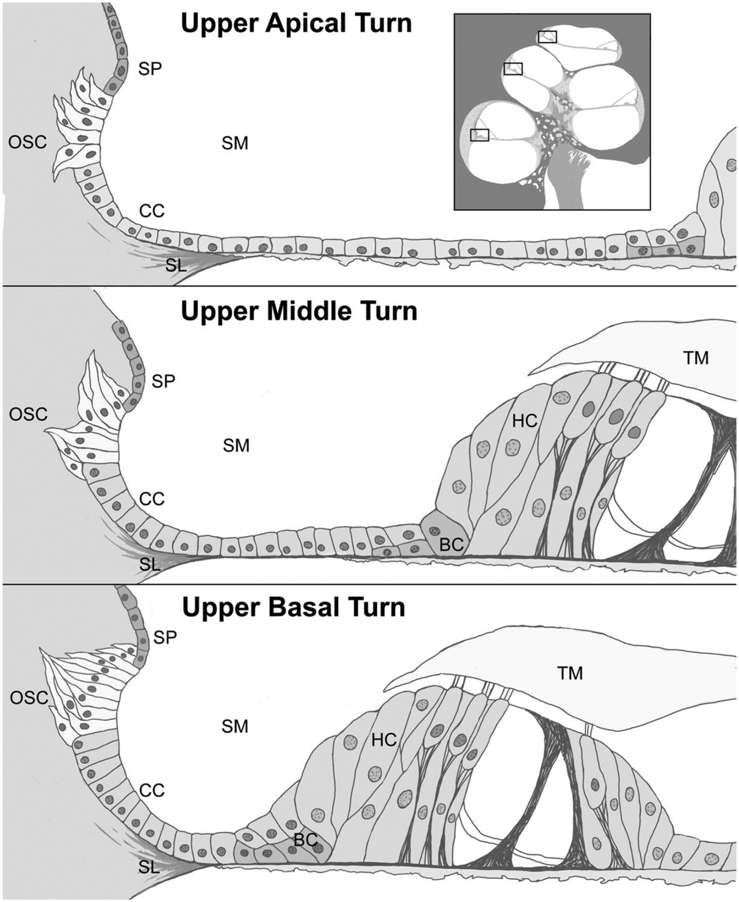
Outer sulcus cells interface with endolymph throughout the cochlea, with greatest contact in the apical turn. Mean outer sulcus cell counts in the upper apical turn (8.82) were generally smaller (all p < 0.05) than those of the upper basal (17.71), lower middle (18.99) upper middle (18.23), and lower apical (16.42) turns. Mean outer sulcus cell counts were higher (p < 0.05) in normal controls (20.1) than in diseased specimens (15.29). There was a significant correlation between mean cell counts and tonotopically expected hearing thresholds in the upper basal (r = -0.662, p = 0.0001), lower middle (r = -0.565, p = 0.0017), and upper middle (r = -0.507, p = 0.0136) regions. Other differences in cell morphology, distribution, or relationship with Claudius cells were not appreciated between normal and diseased specimens. Menière's specimens had no apparent unique features in the cochlear apex. Immunofluorescence staining demonstrated outer sulcus cells extending into the spiral ligament in bundles forming tapering processes which differed between the cochlear turns in morphology.
Outer sulcus cells vary throughout the cochlear turns and correlate with hearing status, but not in a manner specific to the underlying diagnoses of Menière's disease or presbycusis.
人们假设人耳蜗的不同区域以及疾病和正常样本之间的外沟细胞特征和分布存在差异。
外沟细胞在外耳液的动态平衡中发挥作用。然而,它们在人类内耳中的解剖结构和分布尚未得到很好的描述。
通过光镜检查了听力正常(n=10)、梅尼埃病(n=10)、平型听力曲线的老年性聋(n=4)和陡降型听力曲线的老年性聋(n=5)的颞骨标本。在外耳蜗各转定量和定性评估外沟细胞。一个标本用微管蛋白免疫荧光染色,并用共聚焦显微镜成像。
外沟细胞与整个耳蜗的内淋巴相接触,在顶端转接触最大。上尖顶转(8.82)的平均外沟细胞计数通常较小(所有 p<0.05)比上基底(17.71)、中下(18.99)、上中(18.23)和下尖(16.42)转。正常对照组(20.1)的平均外沟细胞计数高于疾病组(15.29)(所有 p<0.05)。在上基底(r=-0.662,p=0.0001)、中下(r=-0.565,p=0.0017)和上中(r=-0.507,p=0.0136)区域,平均细胞计数与音调预期听力阈值之间存在显著相关性。在正常和疾病标本之间,没有观察到外沟细胞形态、分布或与 Claudius 细胞关系的其他差异。梅尼埃病标本在耳蜗顶端没有明显的独特特征。免疫荧光染色显示,外沟细胞延伸到螺旋韧带中,形成束状,逐渐变细的过程,在形态上与耳蜗转不同。
外沟细胞在外耳蜗的不同转中存在差异,并与听力状况相关,但与梅尼埃病或老年性聋的潜在诊断无关。