Department of Chemistry, University of Michigan , Ann Arbor, Michigan 48109, United States.
Langmuir. 2014 Jan 28;30(3):823-31. doi: 10.1021/la404055a. Epub 2014 Jan 16.
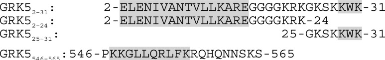
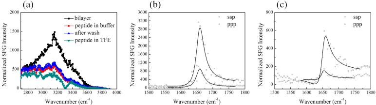
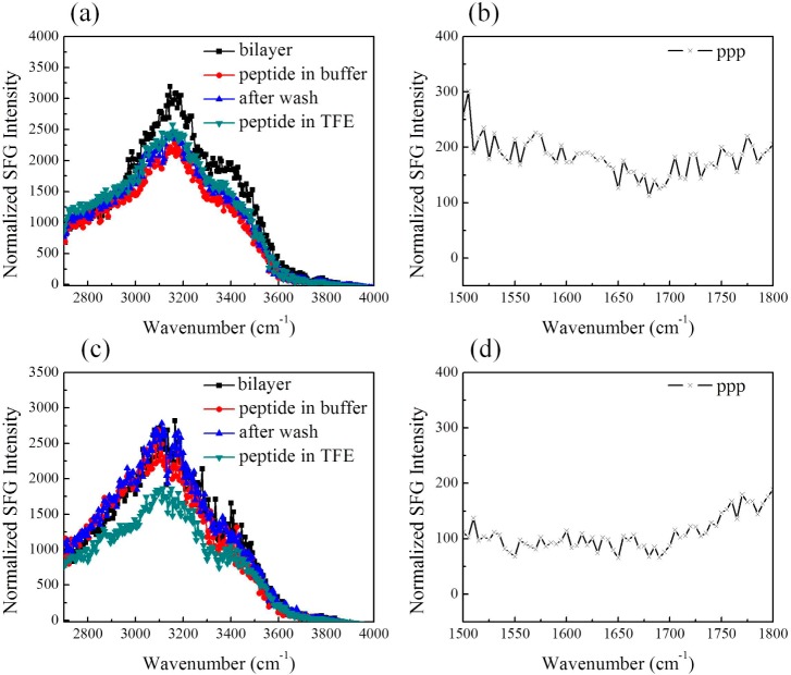
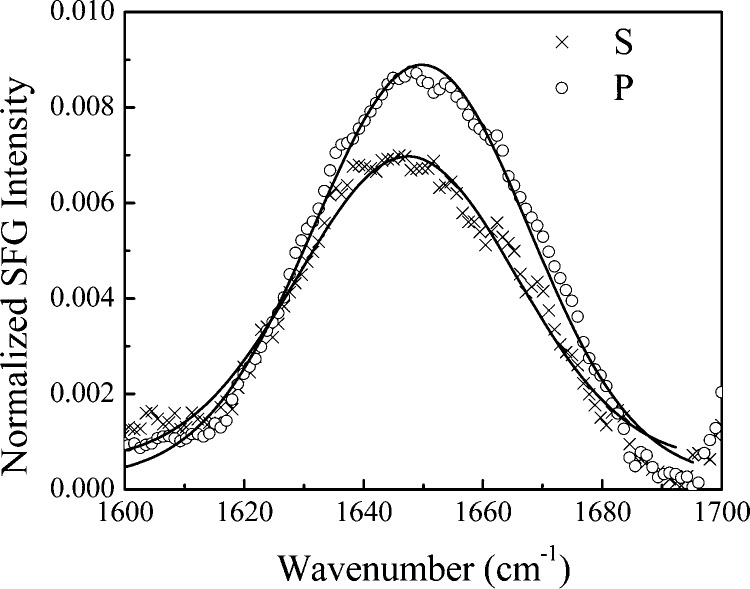
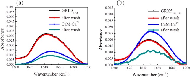
G protein-coupled receptor kinase 5 (GRK5) is thought to associate with membranes in part via N- and C-terminal segments that are typically disordered in available high-resolution crystal structures. Herein we investigate the interactions of these regions with model cell membrane using combined sum frequency generation (SFG) vibrational spectroscopy and attenuated total reflectance-Fourier transform infrared (ATR-FTIR) spectroscopy. It was found that both regions associate with POPC lipid bilayers but adopt different structures when doing so: GRK5 residues 2-31 (GRK5(2-31)) was in random coil whereas GRK5(546-565) was partially helical. When the subphase for the GRK5(2-31) peptide was changed to 40% TFE/60% 10 mM phosphate pH 7.4 buffer, a large change in the SFG amide I signal indicated that GRK5(2-31) became partially helical. By inspecting the membrane behavior of two different segments of GRK5(2-31), namely, GRK5(2-24) and GRK5(25-31), we found that residues 25-31 are responsible for membrane binding, whereas the helical character is imparted by residues 2-24. With SFG, we deduced that the orientation angle of the helical segment of GRK5(2-31) is 46 ± 1° relative to the surface normal in 40% TFE/60% 10 mM phosphate pH = 7.4 buffer but increases to 78 ± 11° with higher ionic strength. We also investigated the effect of PIP2 in the model membrane and concluded that the POPC:PIP2 (9:1) lipid bilayer did not change the behavior of either peptide compared to a pure POPC lipid bilayer. With ATR-FTIR, we also found that Ca(2+)·calmodulin is able to extract both peptides from the POPC lipid bilayer, consistent with the role of this protein in disrupting GRK5 interactions with the plasma membrane in cells.
G 蛋白偶联受体激酶 5(GRK5)被认为通过通常在可用高分辨率晶体结构中无序的 N 端和 C 端片段与膜结合。在此,我们使用和频产生(SFG)振动光谱和衰减全反射-傅里叶变换红外(ATR-FTIR)光谱结合研究了这些区域与模型细胞膜的相互作用。结果发现,这两个区域都与 POPC 脂质双层结合,但结合方式不同:GRK5 残基 2-31(GRK5(2-31))呈无规卷曲,而 GRK5(546-565)呈部分螺旋状。当 GRK5(2-31)肽的亚相被改变为 40%TFE/60%10mM 磷酸盐 pH7.4 缓冲液时,SFG 酰胺 I 信号的大幅变化表明 GRK5(2-31)部分呈螺旋状。通过检查 GRK5(2-31)的两个不同片段,即 GRK5(2-24)和 GRK5(25-31)的膜行为,我们发现残基 25-31 负责膜结合,而螺旋特征则由残基 2-24 赋予。通过 SFG,我们推断 GRK5(2-31)的螺旋段相对于 40%TFE/60%10mM 磷酸盐 pH=7.4 缓冲液中的表面法线的取向角为 46±1°,但在较高离子强度下增加到 78±11°。我们还研究了 PIP2 在模型膜中的影响,并得出结论,与纯 POPC 脂质双层相比,POPC:PIP2(9:1)脂质双层不会改变两种肽的行为。通过 ATR-FTIR,我们还发现 Ca2+-钙调蛋白能够从 POPC 脂质双层中提取这两种肽,这与该蛋白在细胞中破坏 GRK5 与质膜相互作用的作用一致。