Department of Pathology, Gannan Medical University, No, 1, Yixueyuan Road, Ganzhou, Jiangxi 341000, China.
Diagn Pathol. 2014 Jan 20;9:6. doi: 10.1186/1746-1596-9-6.
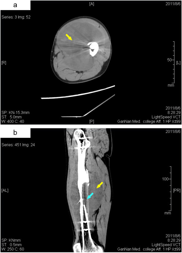
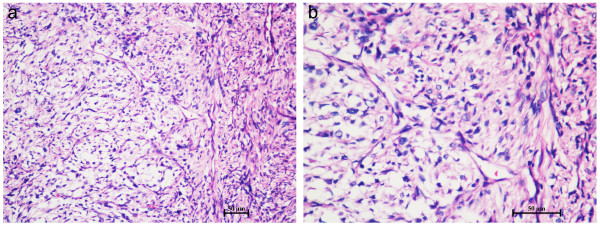
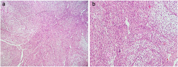
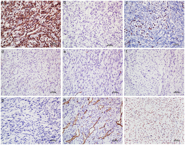
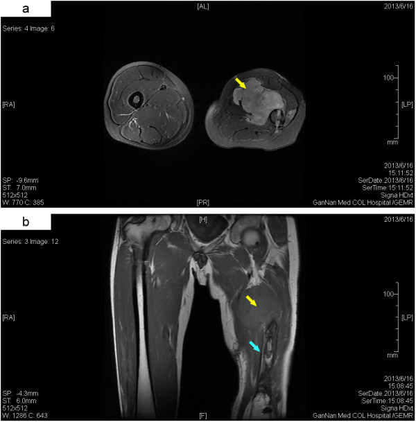
Myxofibrosarcoma is a myxoid variant of malignant fibrous histiocytoma that most commonly involves the extremities of elderly people. However, a primary myxofibrosarcoma with bone invasion in young adults is extremely rare. Herein, we report the case of a 31-year-old male with a gradually enlarging left thigh mass, who had a history of left femur fracture and received an open reduction and internal fixation with titanium alloy plates and screws 33 months previously. Imaging investigations revealed an irregularly shaped soft tissue mass around the left femur shaft and a partial bone defect in the middle one-third of the left femur. Pathological examination of the resected specimen showed a multi-nodular appearance, abundant myxoid matrix and elongated curvilinear capillaries. Immunohistochemical studies revealed that the tumor cells was positive for VIM and MDM2, and was negative for CK, MSA, SMA, DES, S-100 and CD34. Labeling index of Ki-67 was 25%. Based on the morphological finding and immunostaining, it was diagnosed as a low-grade myxofibrosarcoma. The clinical and imaging examinations did not reveal the evidence of a primary cancer elsewhere, and the patient had no personal or family history of malignancy. To our knowledge, this is the first case of a primary myxofibrosarcoma developed following a fracture and metal implantation in young adults.
The virtual slide(s) for this article can be found here: http://www.diagnosticpathology.diagnomx.eu/vs/1745984882113605.
黏液纤维肉瘤是一种黏液样的恶性纤维组织细胞瘤变体,最常累及老年人的四肢。然而,年轻人中伴有骨侵犯的原发性黏液纤维肉瘤极为罕见。在此,我们报告 1 例 31 岁男性,其左大腿逐渐增大肿块,该患者 33 个月前因左股骨骨折接受了切开复位内固定术,使用钛合金板和螺钉固定。影像学检查显示左股骨骨干周围形状不规则的软组织肿块和左股骨中段部分骨缺损。切除标本的病理检查显示多结节外观,丰富的黏液样基质和长曲线毛细血管。免疫组织化学研究显示肿瘤细胞 VIM 和 MDM2 阳性,CK、MSA、SMA、DES、S-100 和 CD34 阴性。Ki-67 标记指数为 25%。基于形态学发现和免疫染色,诊断为低度黏液纤维肉瘤。临床和影像学检查未发现其他部位原发性癌症的证据,患者无恶性肿瘤个人史或家族史。据我们所知,这是首例年轻人骨折和金属植入物后发生的原发性黏液纤维肉瘤。
本文的虚拟幻灯片可以在此处找到:http://www.diagnosticpathology.diagnomx.eu/vs/1745984882113605。