Tsai May-Jywan, Tsai Shen-Kou, Hu Bo-Ruei, Liou Dann-Ying, Huang Shih-Ling, Huang Ming-Chao, Huang Wen-Cheng, Cheng Henrich, Huang Shiang-Suo
Neural Regeneration Laboratory, Center for Neural Regeneration, Department of Neurosurgery, Neurological Institute, Taipei Veterans General Hospital, No, 322, Section 2, Shih-Pai Road, Taipei City, Beitou District 112, Taiwan.
J Biomed Sci. 2014 Jan 22;21(1):5. doi: 10.1186/1423-0127-21-5.
Several lines of evidence have demonstrated that bone marrow-derived mesenchymal stem cells (BM-MSC) release bioactive factors and provide neuroprotection for CNS injury. However, it remains elusive whether BM-MSC derived from healthy donors or stroke patients provides equal therapeutic potential. The present work aims to characterize BM-MSC prepared from normal healthy rats (NormBM-MSC) and cerebral ischemia rats (IschBM-MSC), and examine the effects of their conditioned medium (Cm) on ischemic stroke animal model.
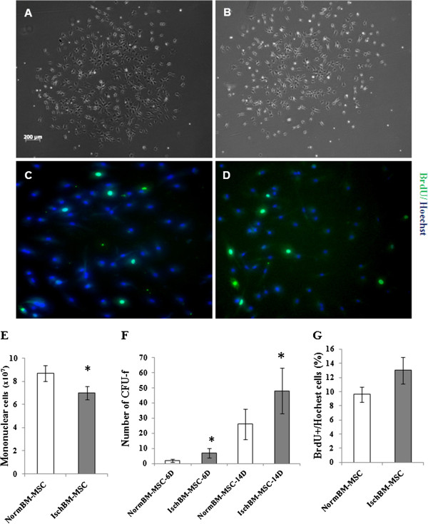
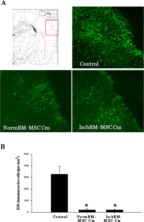
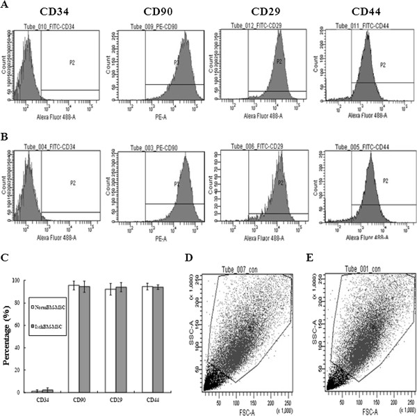
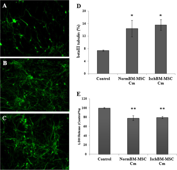
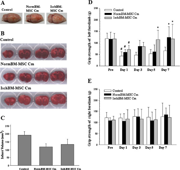
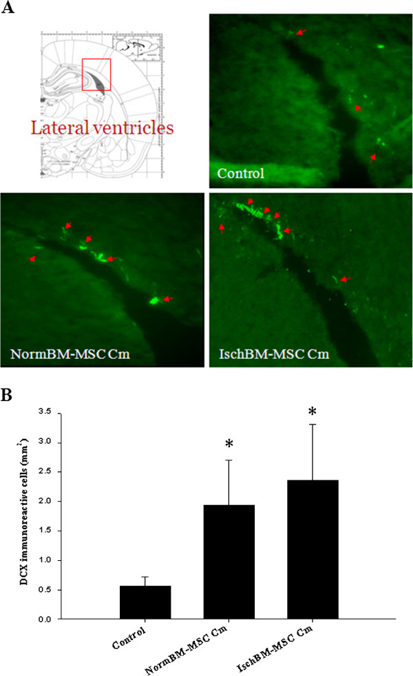
Isolated NormBM-MSC or IschBM-MSC formed fibroblastic like morphology and expressed CD29, CD90 and CD44 but failed to express the hematopoietic marker CD34. The number of colony formation of BM-MSC was more abundant in IschBM-MSC than in NormBM-MSC. This is in contrast to the amount of Ficoll-fractionated mononuclear cells from normal donor and ischemic rats. The effect of cm of BM-MSC was further examined in cultures and in middle cerebral artery occlusion (MCAo) animal model. Both NormBM-MSC Cm and IschBM-MSC Cm effectively increased neuronal connection and survival in mixed neuron-glial cultures. In vivo, intravenous infusion of NormBM-MSC Cm and IschBM-MSC Cm after stroke onset remarkably improved functional recovery. Furthermore, NormBM-MSC Cm and IschBM-MSC Cm increased neurogenesis and attenuated microglia/ macrophage infiltration in MCAo rat brains.
Our data suggest equal effectiveness of BM-MSC Cm derived from ischemic animals or from a normal population. Our results thus revealed the potential of BM-MSC Cm on treatment of ischemic stroke.
多项证据表明,骨髓间充质干细胞(BM-MSC)可释放生物活性因子,并为中枢神经系统损伤提供神经保护。然而,来自健康供体或中风患者的BM-MSC是否具有同等的治疗潜力仍不清楚。本研究旨在对从正常健康大鼠(NormBM-MSC)和脑缺血大鼠(IschBM-MSC)制备的BM-MSC进行表征,并检测其条件培养基(Cm)对缺血性中风动物模型的影响。
分离出的NormBM-MSC或IschBM-MSC呈成纤维细胞样形态,表达CD29、CD90和CD44,但不表达造血标志物CD34。IschBM-MSC中BM-MSC的集落形成数量比NormBM-MSC中更丰富。这与正常供体和缺血大鼠的Ficoll分层单核细胞数量相反。在培养物和大脑中动脉闭塞(MCAo)动物模型中进一步检测了BM-MSC的Cm的作用。NormBM-MSC Cm和IschBM-MSC Cm均能有效增加混合神经元-胶质细胞培养物中的神经元连接和存活。在体内,中风发作后静脉输注NormBM-MSC Cm和IschBM-MSC Cm可显著改善功能恢复。此外,NormBM-MSC Cm和IschBM-MSC Cm可增加MCAo大鼠脑内的神经发生,并减轻小胶质细胞/巨噬细胞浸润。
我们的数据表明,来自缺血动物或正常群体的BM-MSC Cm具有同等效力。因此,我们的结果揭示了BM-MSC Cm在治疗缺血性中风方面的潜力。