Massaro G, Sglavo G, Cavallaro A, Pastore G, Nappi C, Di Carlo C
Department of Obstetrics and Gynecology, University of Naples Federico II, Via Pansini 5, 80131 Naples, Italy.
Case Rep Obstet Gynecol. 2013;2013:764579. doi: 10.1155/2013/764579. Epub 2013 Dec 12.
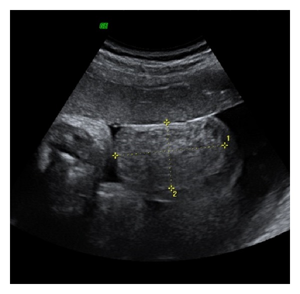
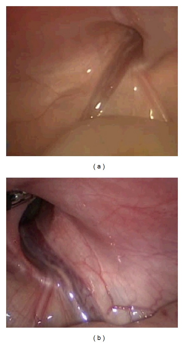
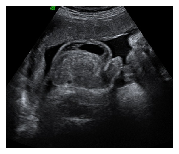
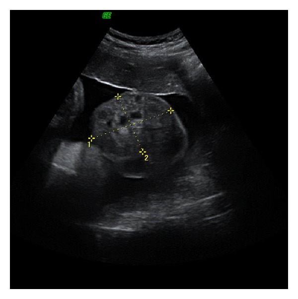
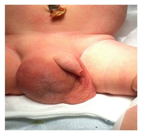
Fetal inguinal scrotal hernia is a rare condition resulting in an abnormal embryonic process of the tunica vaginalis. We report a case of ultrasound prenatal diagnosis of inguinal scrotal hernia associated with contralateral hydrocele in a woman at 37 weeks of gestation, referred to our clinic for a scrotal mass. Differential diagnosis includes hydrocele, teratoma, hemangiomas, solid tumours of testis, bowel herniation, and testicular torsion. Bowel peristalsis is an important ultrasound sign and it allowed us to make diagnosis of inguinal scrotal hernia. Diagnosis was confirmed at birth and a laparoscopic hernia repair was performed without complications on day 10. During surgery, a bilateral defect of canal inguinal was seen and considered as the cause of scrotal inguinal hernia and contralateral hydrocele observed in utero.
胎儿腹股沟阴囊疝是一种罕见的疾病,其导致鞘膜的胚胎发育过程异常。我们报告一例妊娠37周女性的腹股沟阴囊疝合并对侧鞘膜积液的超声产前诊断病例,该女性因阴囊肿物转诊至我院。鉴别诊断包括鞘膜积液、畸胎瘤、血管瘤、睾丸实体瘤、肠疝和睾丸扭转。肠蠕动是一个重要的超声征象,它使我们能够诊断腹股沟阴囊疝。出生时确诊,并在第10天进行了腹腔镜疝修补术,无并发症。手术中发现双侧腹股沟管缺损,并认为这是子宫内观察到的阴囊腹股沟疝和对侧鞘膜积液的原因。