Chen Jing, Liu Wen-Bin, Jia Wei-Dong, Xu Ge-Liang, Ma Jin-Liang, Ren Yun, Chen Hao, Sun Si-Nan, Huang Mei, Li Jian-Sheng
Department of Hepatic Surgery, Anhui Provincial Hospital Affiliated to Anhui Medical University, Hefei, China.
Anhui Province Key Laboratory of Hepatopancreatobiliary Surgery, Anhui Provincial Hospital Affiliated to Anhui Medical University, Hefei, China.
PLoS One. 2014 Jan 21;9(1):e85840. doi: 10.1371/journal.pone.0085840. eCollection 2014.
Nodal, a TGF-β-related embryonic morphogen, is involved in multiple biologic processes. However, the expression of Nodal in hepatocellular carcinoma (HCC) and its correlation with tumor angiogenesis, epithelial-mesenchymal transition, and prognosis is unclear.
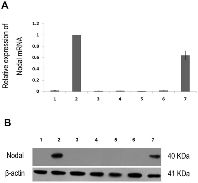
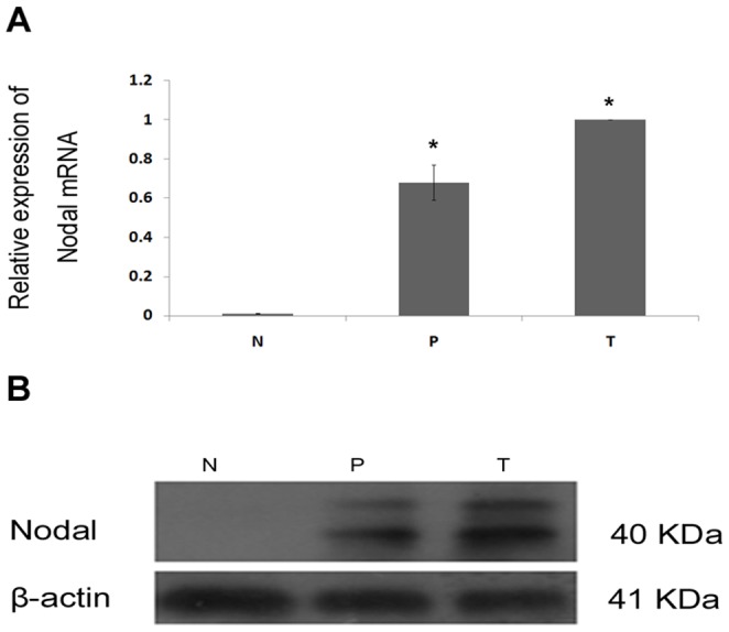
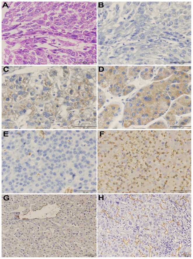
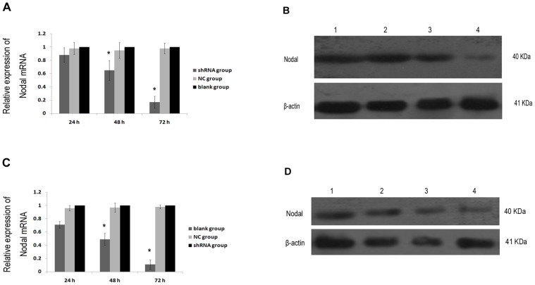
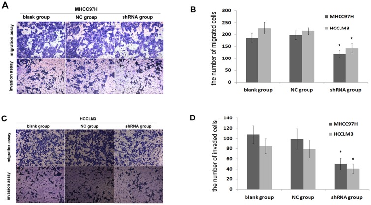
We used real-time PCR and Western blotting to investigate Nodal expression in 6 HCC cell lines and 1 normal liver cell line, 16 pairs of tumor and corresponding paracarcinomatous tissues from HCC patients. Immunohistochemistry was performed to examine Nodal expression in HCC and corresponding paracarcinomatous tissues from 96 patients. CD34 and Vimentin were only examined in HCC tissues of patients mentioned above. Nodal gene was silenced by shRNA in MHCC97H and HCCLM3 cell lines, and cell migration and invasion were detected. Statistical analyses were applied to evaluate the prognostic value and associations of Nodal expression with clinical parameters.
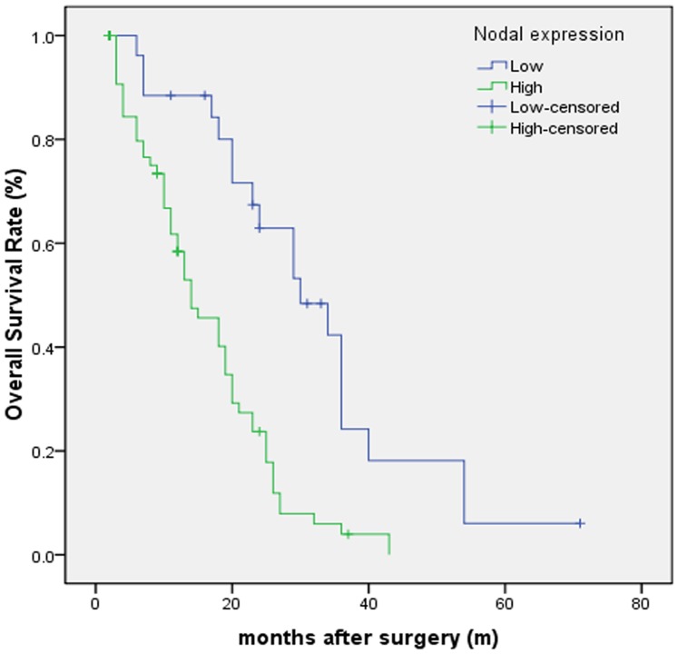
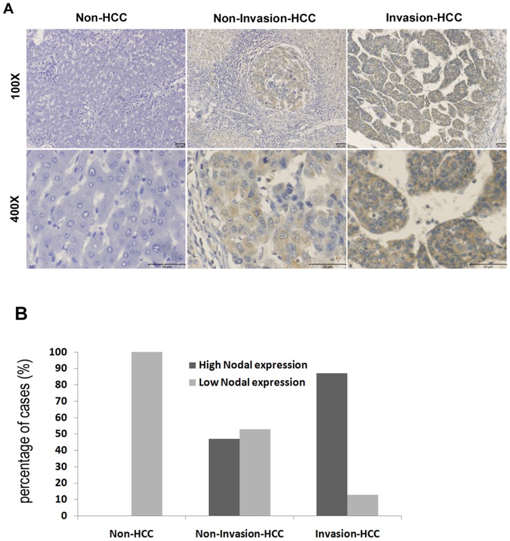
Nodal expression was detected in HCC cell lines with high metastatic potential alone. Nodal expression is up-regulated in HCC tissues compared with paracarcinomatous and normal liver tissues. Nodal protein was expressed in 70 of the 96 (72.9%) HCC tumors, and was associated with vascular invasion (P = 0.000), status of metastasis (P = 0.004), AFP (P = 0.049), ICGR15 (indocyanine green retention rate at 15 min) (P = 0.010) and tumor size (P = 0.000). High Nodal expression was positively correlated with high MVD (microvessal density) (P = 0.006), but not with Vimentin expression (P = 0.053). Significantly fewer migrated and invaded cells were seen in shRNA group compared with blank group and negative control group (P<0.05). High Nodal expression was found to be an independent factor for predicting overall survival of HCC.
Our study demonstrated that Nodal expression is associated with aggressive characteristics of HCC. Its aberrant expression may be a predictive factor of unfavorable prognosis for HCC after surgery.
Nodal是一种与转化生长因子-β(TGF-β)相关的胚胎形态发生素,参与多种生物学过程。然而,Nodal在肝细胞癌(HCC)中的表达及其与肿瘤血管生成、上皮-间质转化和预后的相关性尚不清楚。
我们采用实时定量聚合酶链反应(PCR)和蛋白质免疫印迹法检测6种肝癌细胞系和1种正常肝细胞系、16对肝癌患者肿瘤组织及相应癌旁组织中Nodal的表达。采用免疫组织化学法检测96例肝癌患者肿瘤组织及相应癌旁组织中Nodal的表达。仅对上述患者的肝癌组织检测CD34和波形蛋白。在MHCC97H和HCCLM3细胞系中,通过短发夹RNA(shRNA)沉默Nodal基因,并检测细胞迁移和侵袭能力。应用统计学分析评估Nodal表达的预后价值及其与临床参数的相关性。
仅在具有高转移潜能的肝癌细胞系中检测到Nodal表达。与癌旁组织和正常肝组织相比,肝癌组织中Nodal表达上调。96例肝癌肿瘤组织中有70例(72.9%)表达Nodal蛋白,且与血管侵犯(P = 0.000)、转移状态(P = 0.004)、甲胎蛋白(AFP)(P = 0.049)、15分钟吲哚菁绿潴留率(ICGR15)(P = 0.010)及肿瘤大小(P = 0.000)相关。Nodal高表达与高微血管密度(MVD)呈正相关(P = 0.006),但与波形蛋白表达无关(P = 0.053)。与空白组和阴性对照组相比,shRNA组迁移和侵袭的细胞明显减少(P<0.05)。发现Nodal高表达是预测肝癌患者总生存期的独立因素。
我们的研究表明,Nodal表达与肝癌的侵袭性特征相关。其异常表达可能是肝癌术后预后不良的预测因素。