Vascular and Genomic Center, Changhua Christian Hospital, Changhua, Taiwan ; Institute of Biochemistry and Biotechnology, Chung Shan Medical University, Taichung, Taiwan.
Division of Cardiovascular Center, Department of Internal Medicine, Taichung Veterans General Hospital, Taichung, Taiwan.
PLoS One. 2014 Jan 24;9(1):e71862. doi: 10.1371/journal.pone.0071862. eCollection 2014.
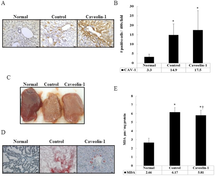
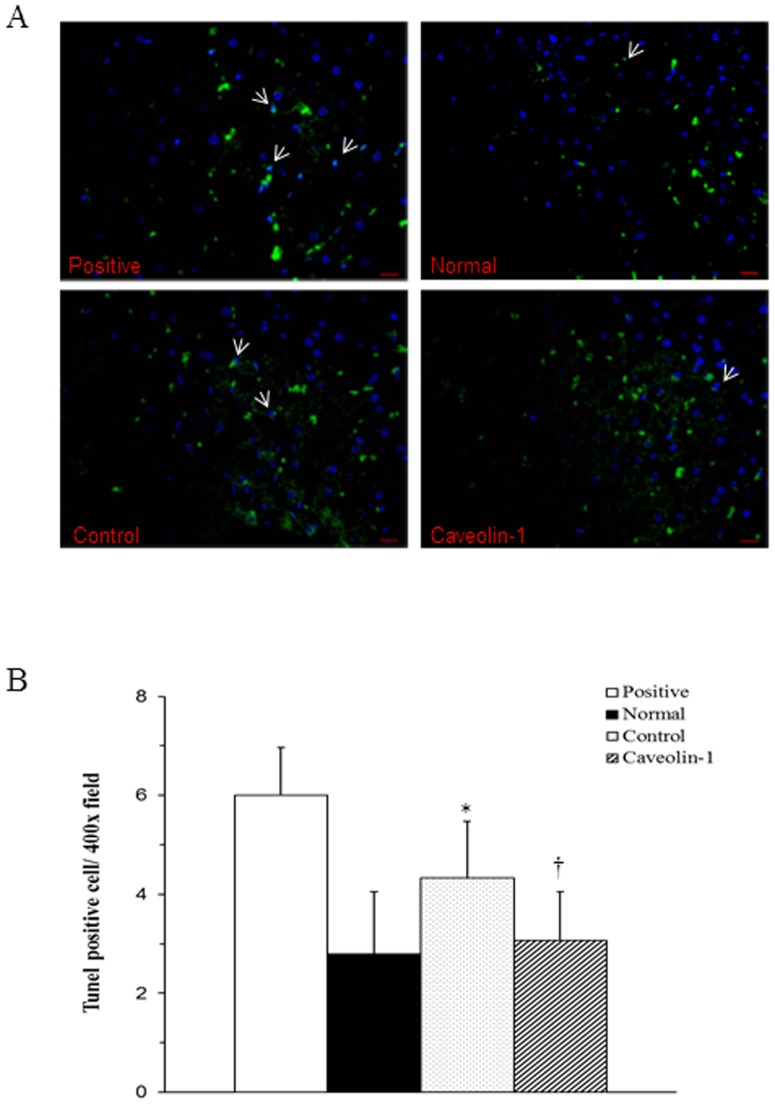
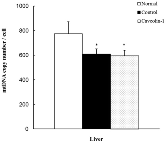
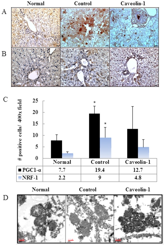
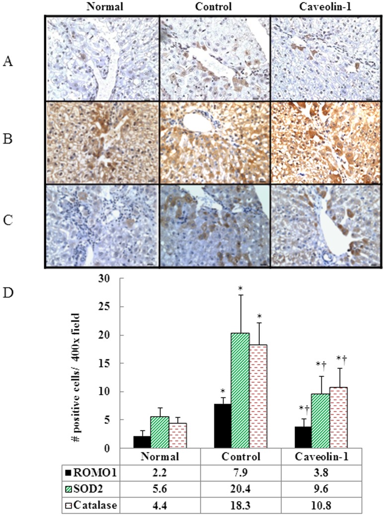
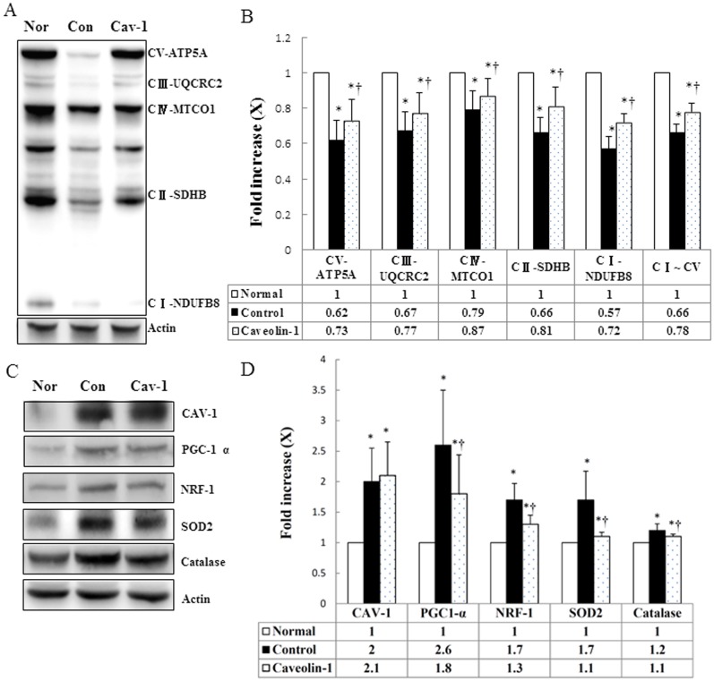
Caveolins are an essential component of cholesterol-rich invaginations of the plasma membrane known as caveolae. These flask-shaped, invaginated structures participate in a number of important cellular processes, including vesicular transport, cholesterol homeostasis, and signal transduction. We investigated the effects of CAV-1 on mitochondrial biogenesis and antioxidant enzymes in hypercholesterolemia-affected target organs. A total of eighteen male New Zealand white rabbits were divided into three groups: a normal-diet group, an untreated hypercholesterolemia-induced group, and a hypercholesterolemia-induced group that received intravenous administration of antennapedia-CAV-1 (AP-CAV-1) peptide every 2 days for 2 weeks. Serum biochemistry, CAV-1 distribution, neutral lipid distribution, mitochondrial morphology, biogenesis-mediated protein content, oxidative stress balance, antioxidant enzyme levels, and apoptotic cell death of liver tissue were analysed. Hepatic and circulating cholesterol and low-density lipoprotein cholesterol (LDL-C) levels differed significantly between the three groups (P<0.05). Immunohistochemical staining intensity of CAV-1 was greater in AP-CAV-1-treated rabbits than in untreated rabbits, especially in the vicinity of the liver vasculature. The high levels of neutral lipids, malondialdehyde, peroxisome proliferator-activated receptor-γ coactive 1α (PGC-1α), and nuclear respiratory factor-1 (NRF-1) seen in untreated hypercholesteremic animals were attenuated by administration of AP-CAV-1 (P<0.05). In addition, mitochondria in animals that received treatment exhibited darker electron-dense matrix and integrated cristae. Furthermore, the levels of ROS modulator 1 (Romo1) and superoxide dismutase (SOD)-2, as well as catalase activity were significantly lower in CAV-1-treated hypercholesterolemic rabbits (P<0.05). AP-CAV-1 treatment also restored mitochondrial respiratory chain subunit protein content (OXPHOS complexes I-V), thereby preserving mitochondrial function (P<0.05). Furthermore, AP-CAV-1 treatment significantly suppressed apoptotic cell death, as evidenced by a reduction in the number of TUNEL-positive cells. Our results indirectly indicate that CAV-1 mediates the negative effects of PGC-1α on hepatic mitochondrial respiratory chain function, promotes the antioxidant enzyme defence system, and maintains mitochondrial biogenesis.
窖蛋白是富含胆固醇的质膜内陷的重要组成部分,称为小窝。这些瓶状的内陷结构参与了许多重要的细胞过程,包括囊泡运输、胆固醇稳态和信号转导。我们研究了 CAV-1 对受高胆固醇影响的靶器官中线粒体生物发生和抗氧化酶的影响。总共 18 只雄性新西兰白兔被分为三组:正常饮食组、未治疗的高胆固醇血症诱导组和每 2 天静脉注射 Antennapedia-CAV-1(AP-CAV-1)肽 2 周的高胆固醇血症诱导组。分析了血清生化、CAV-1 分布、中性脂质分布、线粒体形态、生物发生介导的蛋白质含量、氧化应激平衡、抗氧化酶水平和肝组织的凋亡细胞死亡。三组间肝组织和循环胆固醇及低密度脂蛋白胆固醇(LDL-C)水平差异有统计学意义(P<0.05)。AP-CAV-1 治疗兔的 CAV-1 免疫组化染色强度明显高于未治疗兔,尤其是在肝血管附近。未治疗的高胆固醇血症动物中高水平的中性脂质、丙二醛、过氧化物酶体增殖物激活受体-γ 共激活因子 1α(PGC-1α)和核呼吸因子-1(NRF-1)被 AP-CAV-1 减弱(P<0.05)。此外,接受治疗的动物的线粒体显示出更深的电子致密基质和整合嵴。此外,ROS 调节剂 1(Romo1)和超氧化物歧化酶(SOD)-2 以及过氧化氢酶活性在 CAV-1 治疗的高胆固醇血症兔中显著降低(P<0.05)。AP-CAV-1 治疗还恢复了线粒体呼吸链亚基蛋白含量(OXPHOS 复合物 I-V),从而维持了线粒体功能(P<0.05)。此外,AP-CAV-1 治疗显著抑制了凋亡细胞死亡,TUNEL 阳性细胞数量减少。我们的结果间接表明,CAV-1 介导 PGC-1α 对肝线粒体呼吸链功能的负向影响,促进抗氧化酶防御系统,并维持线粒体生物发生。