Uçeyler Nurcan, Homola György A, Guerrero González Hans, Kramer Daniela, Wanner Christoph, Weidemann Frank, Solymosi László, Sommer Claudia
Department of Neurology, University of Würzburg, Würzburg, Germany ; Würzburg Fabry Center for Interdisciplinary Therapy (FAZIT), University of Würzburg, Würzburg, Germany.
Department of Neuroradiology, University of Würzburg, Würzburg, Germany.
PLoS One. 2014 Jan 27;9(1):e87054. doi: 10.1371/journal.pone.0087054. eCollection 2014.
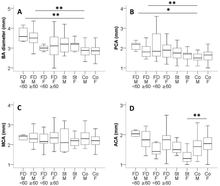
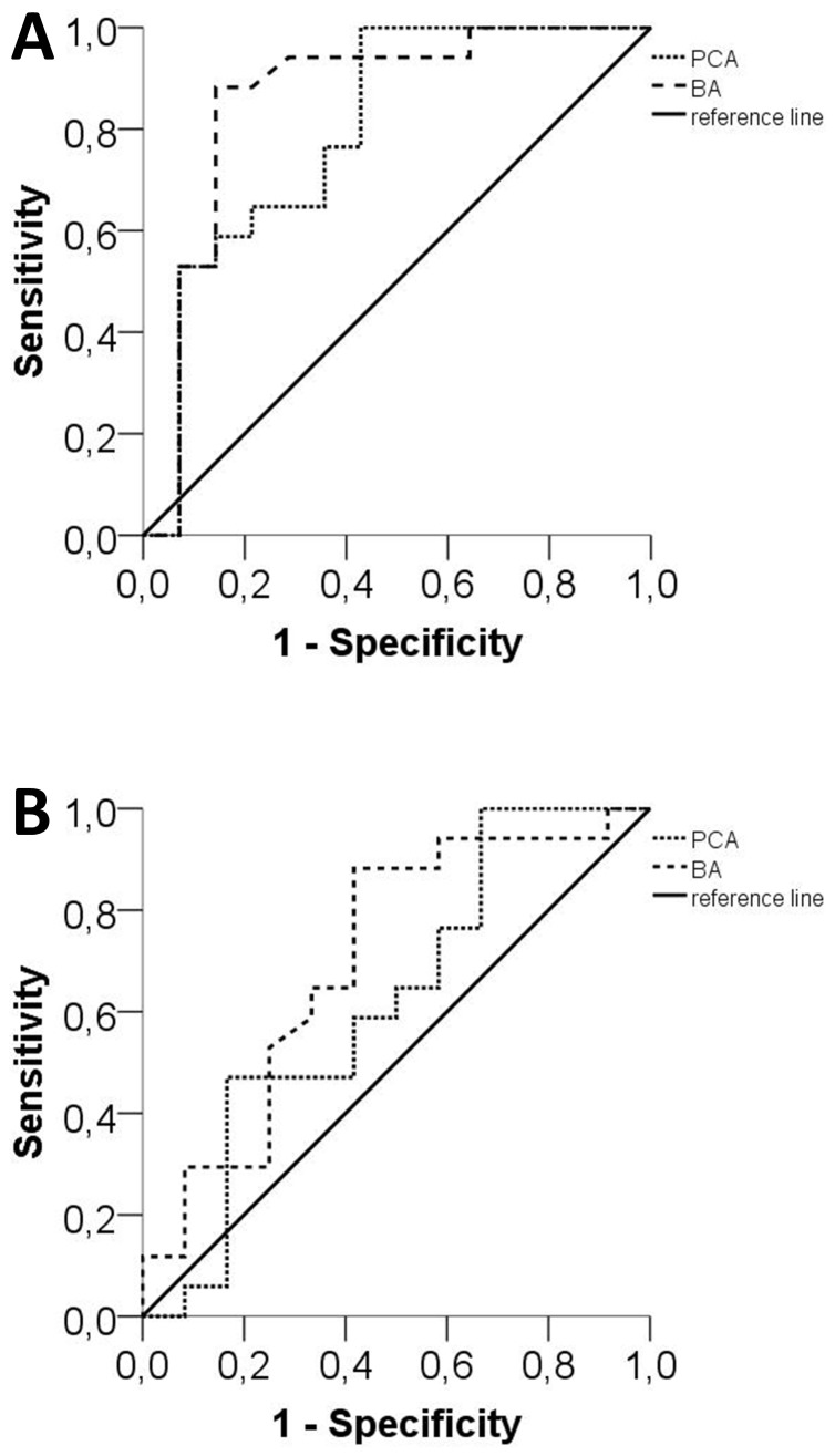
A high load of white matter lesions and enlarged basilar arteries have been shown in selected patients with Fabry disease, a disorder associated with an increased stroke risk. We studied a large cohort of patients with Fabry disease to differentially investigate white matter lesion load and cerebral artery diameters. We retrospectively analyzed cranial magnetic resonance imaging scans of 87 consecutive Fabry patients, 20 patients with ischemic stroke, and 36 controls. We determined the white matter lesion load applying the Fazekas score on fluid-attenuated inversion recovery sequences and measured the diameters of cerebral arteries on 3D-reconstructions of the time-of-flight-MR-angiography scans. Data of different Fabry patient subgroups (males-females; normal-impaired renal function) were compared with data of patients with stroke and controls. A history of stroke or transient ischemic attacks was present in 4/30 males (13%) and 5/57 (9%) females with Fabry disease, all in the anterior circulation. Only one man with Fabry disease showed confluent cerebral white matter lesions in the Fazekas score assessment (1%). Male Fabry patients had a larger basilar artery (p<0.01) and posterior cerebral artery diameter (p<0.05) compared to male controls. This was independent of disease severity as measured by renal function and did not lead to changes in arterial blood flow properties. A basilar artery diameter of >3.2 mm distinguished between men with Fabry disease and controls (sensitivity: 87%, specificity: 86%, p<0.001), but not from stroke patients. Enlarged arterial diameters of the posterior circulation are present only in men with Fabry disease independent of disease severity.
在部分法布里病患者中已发现高负荷的白质病变和基底动脉增粗,法布里病是一种与中风风险增加相关的疾病。我们研究了一大群法布里病患者,以分别调查白质病变负荷和脑动脉直径。我们回顾性分析了87例连续的法布里病患者、20例缺血性中风患者和36例对照者的头颅磁共振成像扫描。我们在液体衰减反转恢复序列上应用 Fazekas 评分确定白质病变负荷,并在时间飞跃磁共振血管造影扫描的三维重建上测量脑动脉直径。将不同法布里病患者亚组(男性 - 女性;肾功能正常 - 受损)的数据与中风患者和对照者的数据进行比较。在患有法布里病的4/30名男性(13%)和5/57名女性(9%)中存在中风或短暂性脑缺血发作史,均在前循环。在 Fazekas 评分评估中,只有一名法布里病男性显示融合性脑白质病变(1%)。与男性对照相比,男性法布里病患者的基底动脉(p<0.01)和大脑后动脉直径更大(p<0.05)。这与通过肾功能衡量的疾病严重程度无关,并且未导致动脉血流特性改变。基底动脉直径>3.2 mm可区分法布里病男性和对照者(敏感性:87%,特异性:86%,p<0.001),但不能区分中风患者。后循环动脉直径增大仅存在于法布里病男性中,与疾病严重程度无关。