Department of Radiology, Seoul National University College of Medicine, Seoul 110-744, Korea.
Department of Neurosurgery, Seoul National University College of Medicine, Seoul 110-744, Korea.
Korean J Radiol. 2014 Jan-Feb;15(1):161-8. doi: 10.3348/kjr.2014.15.1.161. Epub 2014 Jan 8.
The aim of this study was to determine the interobserver and intermodality agreement in the interpretation of time-of-flight (TOF) MR angiography (MRA) for the follow-up of coiled intracranial aneurysms with the Enterprise stent.
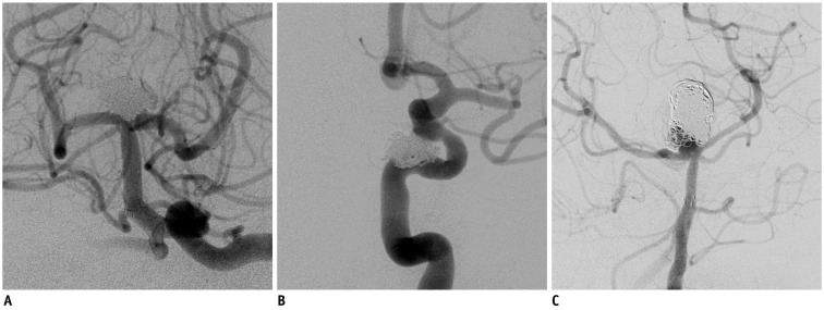
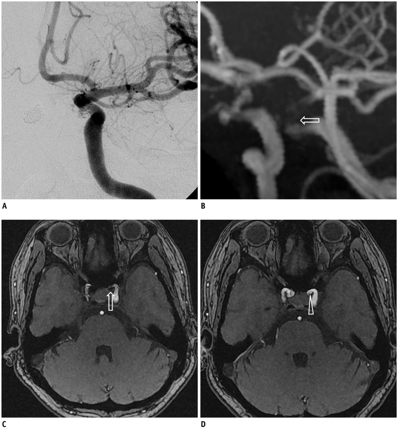
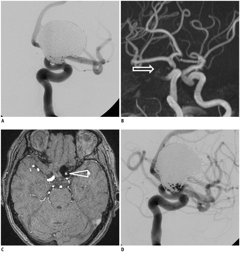
Two experienced neurointerventionists independently reviewed the follow-up MRA studies of 40 consecutive patients with 44 coiled aneurysms. All aneurysms were treated with assistance from the Enterprise stent and the radiologic follow-up intervals were greater than 6 months after the endovascular therapy. Digital subtraction angiography (DSA) served as the reference standard. The degree of aneurysm occlusion was determined by an evaluation of the maximal intensity projection (MIP) and source images (SI) of the TOF MRA. The capability of the TOF MRA to depict the residual flow within the coiled aneurysms and the stented parent arteries was compared with that of the DSA.
DSA showed stable occlusions in 25 aneurysms, minor recanalization in 8, and major recanalization in 11. Comparisons between the TOF MRA and conventional angiography showed that the MIP plus SI had almost perfect agreement (κ = 0.892, range 0.767 to 1.000) and had better agreement than with the MIP images only (κ = 0.598, range 0.370 to 0.826). In-stent stenosis of more than 33% was observed in 5 cases. Both MIP and SI of the MRA showed poor depiction of in-stent stenosis compared with the DSA.
TOF MRA seemed to be reliable in screening for aneurysm recurrence after coil embolization with Enterprise stent assistance, especially in the evaluation of the SI, in addition to MIP images in the TOF MRA.
本研究旨在评估时间飞跃(TOF)磁共振血管造影(MRA)在Enterprise 支架辅助治疗颅内圈闭动脉瘤随访中的观察者间和模态间一致性。
两位有经验的神经介入医师独立分析了 40 例 44 个颅内圈闭动脉瘤患者的连续随访 MRA 资料。所有动脉瘤均在 Enterprise 支架的辅助下进行治疗,血管内治疗结束后随访时间均大于 6 个月。数字减影血管造影(DSA)作为参考标准。采用最大密度投影(MIP)和源图像(SI)对 TOF MRA 评估动脉瘤的闭塞程度。比较 TOF MRA 显示圈闭动脉瘤和支架内母动脉内残余血流的能力与 DSA 的能力。
DSA 显示 25 个动脉瘤稳定闭塞,8 个动脉瘤轻微再通,11 个动脉瘤完全再通。TOF MRA 与传统血管造影的比较显示,MIP 加 SI 具有几乎完美的一致性(κ=0.892,范围 0.767-1.000),且一致性优于 MIP 图像(κ=0.598,范围 0.370-0.826)。5 例支架内狭窄超过 33%。与 DSA 相比,MRA 的 MIP 和 SI 均不能很好地显示支架内狭窄。
TOF MRA 似乎可用于筛查 Enterprise 支架辅助治疗颅内圈闭动脉瘤栓塞后的复发,特别是在评价 SI 时,除了 TOF MRA 的 MIP 图像外。