Yamauchi Motohiro, Otsuka Kensuke, Kondo Hisayoshi, Hamada Nobuyuki, Tomita Masanori, Takahashi Masayuki, Nakasono Satoshi, Iwasaki Toshiyasu, Yoshida Kazuo
Radiation Safety Research Center, Nuclear Technology Research Laboratory, Central Research Institute of Electric Power Industry (CRIEPI), 2-11-1 Iwado Kita, Komae, Tokyo 201-8511, Japan.
J Radiat Res. 2014 Mar 1;55(2):381-90. doi: 10.1093/jrr/rrt123. Epub 2014 Feb 6.
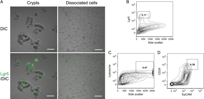
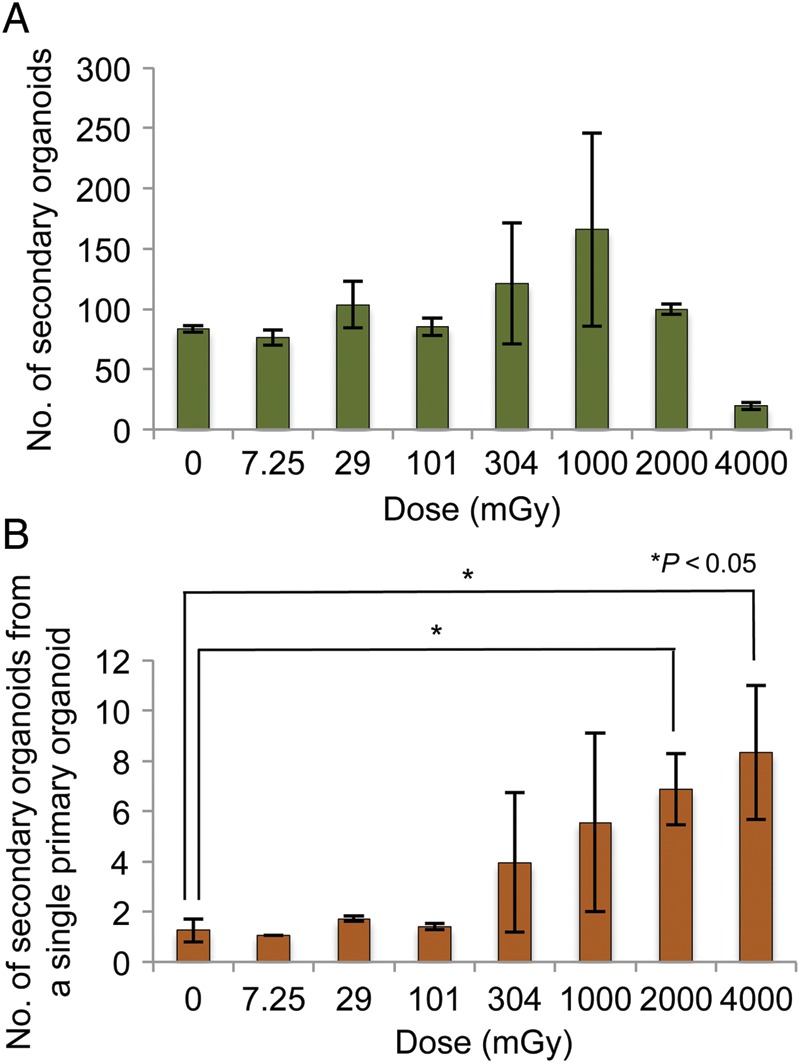
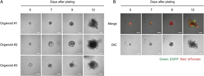
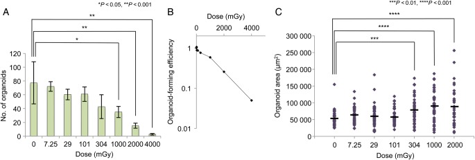
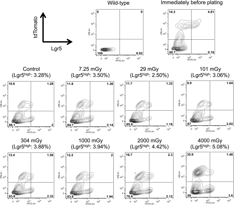
The microcolony assay developed by Withers and Elkind has been a gold standard to assess the surviving fraction of small intestinal stem cells after exposure to high (≥8 Gy) doses of ionizing radiation (IR), but is not applicable in cases of exposure to lower doses. Here, we developed a novel in vitro assay that enables assessment of the surviving fraction of small intestinal stem cells after exposure to lower IR doses. The assay includes in vitro culture of small intestinal stem cells, which allows the stem cells to develop into epithelial organoids containing all four differentiated cell types of the small intestine. We used Lgr5-EGFP-IRES-CreERT2/ROSA26-tdTomato mice to identify Lgr5(+) stem cells and their progeny. Enzymatically dissociated single crypt cells from the duodenum and jejunum of mice were irradiated with 7.25, 29, 101, 304, 1000, 2000 and 4000 mGy of X-rays immediately after plating, and the number of organoids was counted on Day 12. Organoid-forming efficiency of irradiated cells relative to that of unirradiated controls was defined as the surviving fraction of stem cells. We observed a significant decrease in the surviving fraction of stem cells at ≥1000 mGy. Moreover, fluorescence-activated cell sorting analyses and passage of the organoids revealed that proliferation of stem cells surviving IR is significantly potentiated. Together, the present study demonstrates that the in vitro assay is useful for quantitatively assessing the surviving fraction of small intestinal stem cells after exposure to lower doses of IR as compared with previous examinations using the microcolony assay.
由威瑟斯和埃尔金德开发的微集落试验一直是评估小肠干细胞在接受高剂量(≥8 Gy)电离辐射(IR)后存活分数的金标准,但不适用于低剂量照射的情况。在此,我们开发了一种新型体外试验,能够评估小肠干细胞在接受低剂量IR照射后的存活分数。该试验包括小肠干细胞的体外培养,这使得干细胞能够发育成包含小肠所有四种分化细胞类型的上皮类器官。我们使用Lgr5-EGFP-IRES-CreERT2/ROSA26-tdTomato小鼠来鉴定Lgr5(+)干细胞及其后代。从小鼠十二指肠和空肠中酶解分离出的单个隐窝细胞在接种后立即接受7.25、29、101、304、1000、2000和4000 mGy的X射线照射,并在第12天对类器官数量进行计数。将照射细胞相对于未照射对照的类器官形成效率定义为干细胞的存活分数。我们观察到在≥1000 mGy时干细胞的存活分数显著下降。此外,荧光激活细胞分选分析和类器官传代显示,IR后存活的干细胞增殖显著增强。总之,本研究表明,与先前使用微集落试验的检查相比,该体外试验可用于定量评估小肠干细胞在接受低剂量IR照射后的存活分数。