Imaeda Masumi, Ishikawa Hitoshi, Yoshida Yukari, Takahashi Takeo, Ohkubo Yu, Musha Atsushi, Komachi Mayumi, Nakazato Yoichi, Nakano Takashi
Department of Radiation Oncology, Gunma University, Graduate School of Medicine, 3-39-22 Showa, Maebashi, Gunma 371-8511, Japan.
Department of Radiation Oncology, University of Tsukuba, Faculty of Medicine, 1-1-1 Tennodai, Tsukuba, Ibaraki 305-8575, Japan
J Radiat Res. 2014 Jul;55(4):665-73. doi: 10.1093/jrr/rru005. Epub 2014 Feb 23.
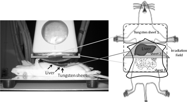
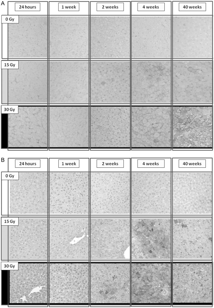
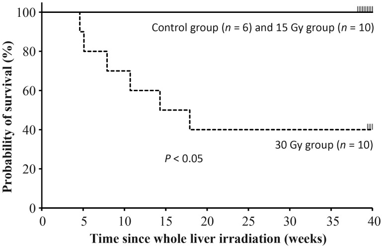
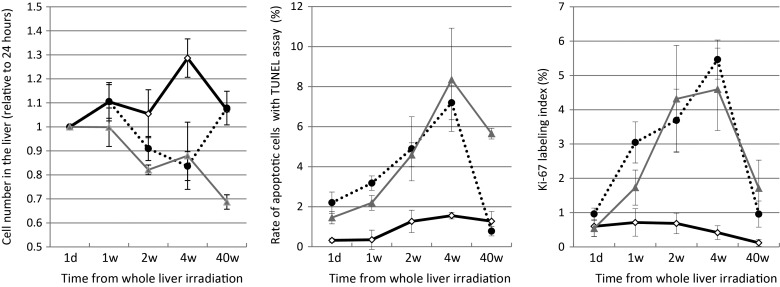
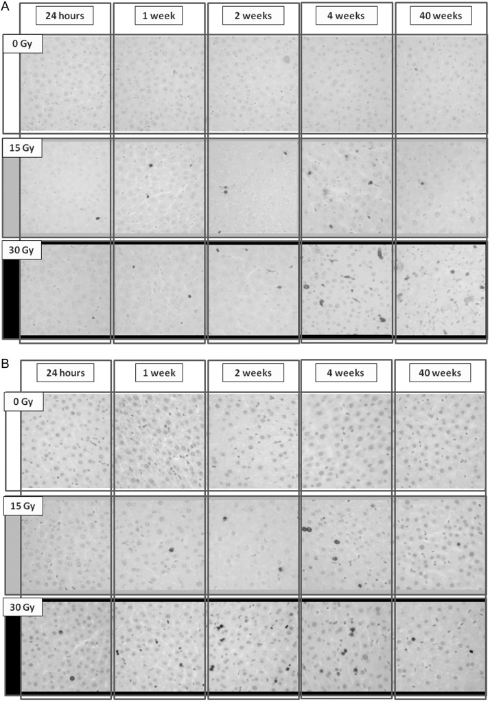
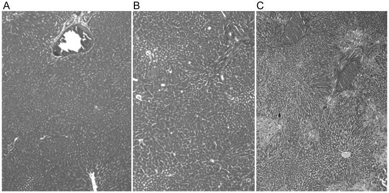
Radiation therapy (RT) has become particularly important recently for treatment of liver tumors, but there are few experimental investigations pertaining to radiation-induced liver injuries over long-term follow-up periods. Thus, the present study examined pathological liver features over a 10-month period using an intraoperative whole-liver irradiation model. Liver function tests were performed in blood samples, whereas cell death, cell proliferation, and fibrotic changes were evaluated pathologically in liver tissues, which were collected from irradiated rats 24 h, 1, 2, 4 and 40 weeks following administration of single irradiation doses of 0 (control), 15 or 30 Gy. The impaired liver function, increased hepatocyte number, and decreased apoptotic cell proportion observed in the 15 Gy group, but not the 30 Gy group, returned to control group levels after 40 weeks; however, the Ki-67 indexes in the 15 Gy group were still higher than those in the control group after 40 weeks. Azan staining showed a fibrotic pattern in the irradiated liver in the 30 Gy group only, but the expression levels of alpha smooth muscle actin (α-SMA) and transforming growth factor-beta 1 (TGF-β1) in both the 15 and 30 Gy groups were significantly higher than those in the control group (P < 0.05). There were differences in the pathological features of the irradiated livers between the 15 Gy and 30 Gy groups, but TGF-β1 and α-SMA expression patterns supported the gradual progression of radiation-induced liver fibrosis in both groups. These findings will be useful in the future development of protective drugs for radiation-induced liver injury.
放射治疗(RT)最近在肝癌治疗中变得尤为重要,但关于长期随访期内辐射诱导肝损伤的实验研究较少。因此,本研究使用术中全肝照射模型,在10个月的时间内检查了肝脏的病理特征。采集了单次照射剂量为0(对照)、15或30 Gy的大鼠在照射后24小时、1、2、4和40周的肝脏组织,对血液样本进行肝功能测试,同时对肝脏组织中的细胞死亡、细胞增殖和纤维化变化进行病理评估。15 Gy组而非30 Gy组出现的肝功能受损、肝细胞数量增加和凋亡细胞比例降低在40周后恢复到对照组水平;然而,40周后15 Gy组的Ki-67指数仍高于对照组。阿赞染色显示仅在30 Gy组的照射肝脏中出现纤维化模式,但15和30 Gy组的α平滑肌肌动蛋白(α-SMA)和转化生长因子-β1(TGF-β1)表达水平均显著高于对照组(P<0.05)。15 Gy组和30 Gy组照射肝脏的病理特征存在差异,但TGF-β1和α-SMA表达模式支持两组辐射诱导肝纤维化的逐渐进展。这些发现将有助于未来开发辐射诱导肝损伤的保护药物。