• 文献检索
  • 文档翻译
  • 深度研究
  • 学术资讯
  • Suppr Zotero 插件Zotero 插件
  • 邀请有礼
  • 套餐&价格
  • 历史记录
应用&插件
Suppr Zotero 插件Zotero 插件浏览器插件Mac 客户端Windows 客户端微信小程序
定价
高级版会员购买积分包购买API积分包
服务
文献检索文档翻译深度研究API 文档MCP 服务
关于我们
关于 Suppr公司介绍联系我们用户协议隐私条款
关注我们

Suppr 超能文献

核心技术专利:CN118964589B侵权必究
粤ICP备2023148730 号-1Suppr @ 2026

文献检索

告别复杂PubMed语法,用中文像聊天一样搜索,搜遍4000万医学文献。AI智能推荐,让科研检索更轻松。

立即免费搜索

文件翻译

保留排版,准确专业,支持PDF/Word/PPT等文件格式,支持 12+语言互译。

免费翻译文档

深度研究

AI帮你快速写综述,25分钟生成高质量综述,智能提取关键信息,辅助科研写作。

立即免费体验

PTRN-1是一种微管负端结合的CAMSAP同源物,可促进秀丽隐杆线虫神经元中的微管功能。

PTRN-1, a microtubule minus end-binding CAMSAP homolog, promotes microtubule function in Caenorhabditis elegans neurons.

作者信息

Richardson Claire E, Spilker Kerri A, Cueva Juan G, Perrino John, Goodman Miriam B, Shen Kang

机构信息

Department of Biology, Stanford University, Stanford, United States.

出版信息

Elife. 2014 Feb 25;3:e01498. doi: 10.7554/eLife.01498.

DOI:10.7554/eLife.01498
PMID:24569477
原文链接:https://pmc.ncbi.nlm.nih.gov/articles/PMC3932522/
Abstract

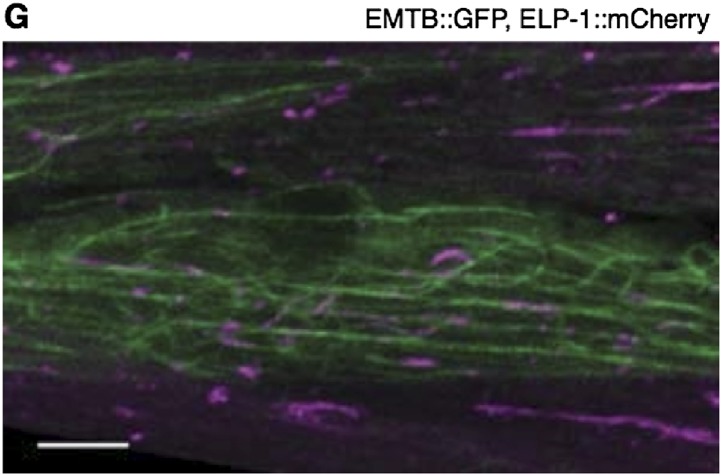
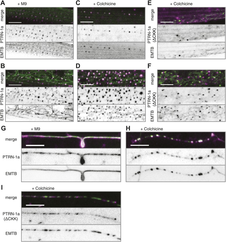
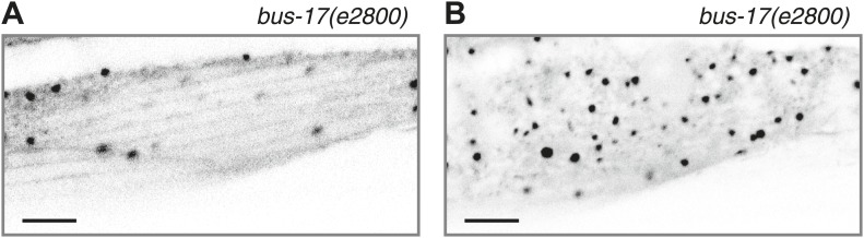
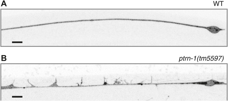
In neuronal processes, microtubules (MTs) provide structural support and serve as tracks for molecular motors. While it is known that neuronal MTs are more stable than MTs in non-neuronal cells, the molecular mechanisms underlying this stability are not fully understood. In this study, we used live fluorescence microscopy to show that the C. elegans CAMSAP protein PTRN-1 localizes to puncta along neuronal processes, stabilizes MT foci, and promotes MT polymerization in neurites. Electron microscopy revealed that ptrn-1 null mutants have fewer MTs and abnormal MT organization in the PLM neuron. Animals grown with a MT depolymerizing drug caused synthetic defects in neurite branching in the absence of ptrn-1 function, indicating that PTRN-1 promotes MT stability. Further, ptrn-1 null mutants exhibited aberrant neurite morphology and synaptic vesicle localization that is partially dependent on dlk-1. Our results suggest that PTRN-1 represents an important mechanism for promoting MT stability in neurons. DOI: http://dx.doi.org/10.7554/eLife.01498.001.

摘要

在神经元突起中,微管(MTs)提供结构支撑并作为分子马达的轨道。虽然已知神经元微管比非神经元细胞中的微管更稳定,但其稳定性背后的分子机制尚未完全了解。在本研究中,我们使用实时荧光显微镜显示秀丽隐杆线虫的CAMSAP蛋白PTRN-1定位于沿神经元突起的斑点,稳定微管焦点,并促进神经突中的微管聚合。电子显微镜显示ptrn-1基因敲除突变体在PLM神经元中的微管较少且微管组织异常。在缺乏ptrn-1功能的情况下,用微管解聚药物培养的动物在神经突分支中导致合成缺陷,表明PTRN-1促进微管稳定性。此外,ptrn-1基因敲除突变体表现出异常的神经突形态和突触小泡定位,这部分依赖于dlk-1。我们的结果表明,PTRN-1代表了促进神经元中微管稳定性的重要机制。DOI:http://dx.doi.org/10.7554/eLife.01498.001。

https://cdn.ncbi.nlm.nih.gov/pmc/blobs/cd3f/3932522/36ee4c472e90/elife01498f008.jpg
https://cdn.ncbi.nlm.nih.gov/pmc/blobs/cd3f/3932522/1fd807ea7680/elife01498f001.jpg
https://cdn.ncbi.nlm.nih.gov/pmc/blobs/cd3f/3932522/20f4b57266cc/elife01498fs001.jpg
https://cdn.ncbi.nlm.nih.gov/pmc/blobs/cd3f/3932522/7a4f309e96b9/elife01498fs002.jpg
https://cdn.ncbi.nlm.nih.gov/pmc/blobs/cd3f/3932522/3922f51410d6/elife01498fs003.jpg
https://cdn.ncbi.nlm.nih.gov/pmc/blobs/cd3f/3932522/576a412fb734/elife01498fs004.jpg
https://cdn.ncbi.nlm.nih.gov/pmc/blobs/cd3f/3932522/b8ea7bb2c0dd/elife01498fs005.jpg
https://cdn.ncbi.nlm.nih.gov/pmc/blobs/cd3f/3932522/999fe06a8c47/elife01498f002.jpg
https://cdn.ncbi.nlm.nih.gov/pmc/blobs/cd3f/3932522/f7ca5c7fb209/elife01498fs006.jpg
https://cdn.ncbi.nlm.nih.gov/pmc/blobs/cd3f/3932522/48cd2d9b28b0/elife01498fs007.jpg
https://cdn.ncbi.nlm.nih.gov/pmc/blobs/cd3f/3932522/79578e196c10/elife01498fs008.jpg
https://cdn.ncbi.nlm.nih.gov/pmc/blobs/cd3f/3932522/2b81d9fd8c0b/elife01498f003.jpg
https://cdn.ncbi.nlm.nih.gov/pmc/blobs/cd3f/3932522/e2e20a86727f/elife01498fs009.jpg
https://cdn.ncbi.nlm.nih.gov/pmc/blobs/cd3f/3932522/06460a97c1d2/elife01498f004.jpg
https://cdn.ncbi.nlm.nih.gov/pmc/blobs/cd3f/3932522/b47319487dfb/elife01498fs010.jpg
https://cdn.ncbi.nlm.nih.gov/pmc/blobs/cd3f/3932522/26d3f05b4a44/elife01498f005.jpg
https://cdn.ncbi.nlm.nih.gov/pmc/blobs/cd3f/3932522/779b07151ae5/elife01498fs011.jpg
https://cdn.ncbi.nlm.nih.gov/pmc/blobs/cd3f/3932522/6096029811f7/elife01498f006.jpg
https://cdn.ncbi.nlm.nih.gov/pmc/blobs/cd3f/3932522/fcc2cd3ea826/elife01498f007.jpg
https://cdn.ncbi.nlm.nih.gov/pmc/blobs/cd3f/3932522/80c81288711d/elife01498fs012.jpg
https://cdn.ncbi.nlm.nih.gov/pmc/blobs/cd3f/3932522/36ee4c472e90/elife01498f008.jpg

相似文献

1
PTRN-1, a microtubule minus end-binding CAMSAP homolog, promotes microtubule function in Caenorhabditis elegans neurons.PTRN-1是一种微管负端结合的CAMSAP同源物,可促进秀丽隐杆线虫神经元中的微管功能。
Elife. 2014 Feb 25;3:e01498. doi: 10.7554/eLife.01498.
2
The Caenorhabditis elegans microtubule minus-end binding homolog PTRN-1 stabilizes synapses and neurites.秀丽隐杆线虫微管负端结合同源物PTRN-1可稳定突触和神经突。
Elife. 2014 Feb 25;3:e01637. doi: 10.7554/eLife.01637.
3
A tubulin-MAPKKK pathway engages tubulin isotype interaction for neuroprotection.一条微管蛋白-促分裂原活化蛋白激酶激酶激酶(MAPKKK)信号通路通过微管蛋白亚型相互作用实现神经保护作用。
Proc Natl Acad Sci U S A. 2025 Aug 26;122(34):e2507208122. doi: 10.1073/pnas.2507208122. Epub 2025 Aug 14.
4
The microtubule minus-end-binding protein patronin/PTRN-1 is required for axon regeneration in C. elegans.微管负端结合蛋白patronin/PTRN-1是秀丽隐杆线虫轴突再生所必需的。
Cell Rep. 2014 Nov 6;9(3):874-83. doi: 10.1016/j.celrep.2014.09.054. Epub 2014 Oct 23.
5
The doublecortin-family kinase ZYG-8DCLK1 regulates microtubule dynamics and motor-driven forces to promote the stability of C. elegans acentrosomal spindles.双皮质素家族激酶 ZYG-8DCLK1 调节微管动力学和马达驱动力以促进线虫无中心体纺锤体的稳定性。
PLoS Genet. 2024 Sep 3;20(9):e1011373. doi: 10.1371/journal.pgen.1011373. eCollection 2024 Sep.
6
Kinesin-1 regulates dendrite microtubule polarity in Caenorhabditis elegans.驱动蛋白-1调节秀丽隐杆线虫中的树突微管极性。
Elife. 2013 Mar 6;2:e00133. doi: 10.7554/eLife.00133.
7
PTRN-1 (CAMSAP) and NOCA-2 (NINEIN) are required for microtubule polarity in Caenorhabditis elegans dendrites.PTRN-1(CAMSAP)和 NOCA-2(NINEIN)对于秀丽隐杆线虫树突中的微管极性是必需的。
PLoS Biol. 2022 Nov 17;20(11):e3001855. doi: 10.1371/journal.pbio.3001855. eCollection 2022 Nov.
8
HIV-1 Exploits CLASP2 To Induce Microtubule Stabilization and Facilitate Virus Trafficking to the Nucleus.HIV-1 利用 CLASP2 诱导微管稳定并促进病毒向核内运输。
J Virol. 2020 Jul 1;94(14). doi: 10.1128/JVI.00404-20.
9
Dynamic microtubules drive circuit rewiring in the absence of neurite remodeling.动态微管在不存在神经突重塑的情况下驱动回路重新布线。
Curr Biol. 2015 Jun 15;25(12):1594-605. doi: 10.1016/j.cub.2015.04.061. Epub 2015 Jun 4.
10
PTRN-1/CAMSAP promotes CYK-1/formin-dependent actin polymerization during endocytic recycling.PTRN-1/CAMSAP 促进内吞循环过程中 CYK-1/formin 依赖性肌动蛋白聚合。
EMBO J. 2018 May 2;37(9). doi: 10.15252/embj.201798556. Epub 2018 Mar 22.

引用本文的文献

1
Announcing the Biomedical Data Translator: Initial Public Release.宣布生物医学数据翻译器:首次公开发布。
Clin Transl Sci. 2025 Jul;18(7):e70284. doi: 10.1111/cts.70284.
2
Dopey-dependent regulation of extracellular vesicles maintains neuronal morphology.呆小蛋白依赖性调节细胞外囊泡维持神经元形态。
Curr Biol. 2024 Nov 4;34(21):4920-4933.e11. doi: 10.1016/j.cub.2024.09.018. Epub 2024 Oct 7.
3
C. elegans touch receptor neurons direct mechanosensory complex organization via repurposing conserved basal lamina proteins.秀丽隐杆线虫触觉受体神经元通过重新利用保守的基底膜蛋白来指导机械感觉复合物的组织。

本文引用的文献

1
Loss of MEC-17 leads to microtubule instability and axonal degeneration.MEC-17 的缺失导致微管不稳定和轴突退化。
Cell Rep. 2014 Jan 16;6(1):93-103. doi: 10.1016/j.celrep.2013.12.004. Epub 2013 Dec 27.
2
Microtubules that form the stationary lattice of muscle fibers are dynamic and nucleated at Golgi elements.形成肌肉纤维固定晶格的微管是动态的,并在高尔基元件处成核。
J Cell Biol. 2013 Oct 28;203(2):205-13. doi: 10.1083/jcb.201304063. Epub 2013 Oct 21.
3
Statistical tests for measures of colocalization in biological microscopy.
Curr Biol. 2024 Jul 22;34(14):3133-3151.e10. doi: 10.1016/j.cub.2024.06.013. Epub 2024 Jul 3.
4
Dopey-dependent regulation of extracellular vesicles maintains neuronal morphology.依赖多配体蛋白聚糖的细胞外囊泡调控维持神经元形态。
bioRxiv. 2024 May 8:2024.05.07.591898. doi: 10.1101/2024.05.07.591898.
5
The outer kinetochore components KNL-1 and Ndc80 complex regulate axon and neuronal cell body positioning in the nervous system.外中心体组件 KNL-1 和 Ndc80 复合物调节神经系统中的轴突和神经元细胞体定位。
Mol Biol Cell. 2024 Jun 1;35(6):ar83. doi: 10.1091/mbc.E23-08-0325. Epub 2024 Apr 24.
6
The response of Dual-leucine zipper kinase (DLK) to nocodazole: Evidence for a homeostatic cytoskeletal repair mechanism.双亮氨酸拉链激酶(DLK)对诺考达唑的反应:细胞骨架稳态修复机制的证据。
PLoS One. 2024 Apr 4;19(4):e0300539. doi: 10.1371/journal.pone.0300539. eCollection 2024.
7
Patronin regulates presynaptic microtubule organization and neuromuscular junction development in .Patronin调节线虫中的突触前微管组织和神经肌肉接头发育。
iScience. 2024 Jan 18;27(2):108944. doi: 10.1016/j.isci.2024.108944. eCollection 2024 Feb 16.
8
Patronin/CAMSAP promotes reactivation and regeneration of Drosophila quiescent neural stem cells.Patronin/CAMSAP 促进果蝇静止神经干细胞的再激活和再生。
EMBO Rep. 2023 Sep 6;24(9):e56624. doi: 10.15252/embr.202256624. Epub 2023 Jul 13.
9
Lipid droplets and peroxisomes are co-regulated to drive lifespan extension in response to mono-unsaturated fatty acids.脂滴和过氧化物酶体协同调节以响应单不饱和脂肪酸延长寿命。
Nat Cell Biol. 2023 May;25(5):672-684. doi: 10.1038/s41556-023-01136-6. Epub 2023 May 1.
10
Non-coding ribonucleic acid-mediated upregulation leads to poor prognosis with suppressed immune infiltration in liver hepatocellular carcinoma.非编码核糖核酸介导的上调导致肝细胞癌预后不良并伴有免疫浸润受抑制。
Front Genet. 2022 Sep 21;13:916847. doi: 10.3389/fgene.2022.916847. eCollection 2022.
生物显微镜共定位测量的统计检验。
J Microsc. 2013 Dec;252(3):295-302. doi: 10.1111/jmi.12093. Epub 2013 Oct 10.
4
Patronin mediates a switch from kinesin-13-dependent poleward flux to anaphase B spindle elongation.伴侣蛋白介导了从依赖于驱动蛋白-13 的极向运输到后期 B 纺锤体伸长的转换。
J Cell Biol. 2013 Oct 14;203(1):35-46. doi: 10.1083/jcb.201306001. Epub 2013 Oct 7.
5
The DLK signalling pathway--a double-edged sword in neural development and regeneration.DLK 信号通路——神经发育和再生中的双刃剑。
EMBO Rep. 2013 Jul;14(7):605-14. doi: 10.1038/embor.2013.64. Epub 2013 May 17.
6
Kinesin-1 regulates dendrite microtubule polarity in Caenorhabditis elegans.驱动蛋白-1调节秀丽隐杆线虫中的树突微管极性。
Elife. 2013 Mar 6;2:e00133. doi: 10.7554/eLife.00133.
7
A dominant mutation in mec-7/β-tubulin affects axon development and regeneration in Caenorhabditis elegans neurons.MEC-7/β-微管蛋白的显性突变影响秀丽隐杆线虫神经元的轴突发育和再生。
Mol Biol Cell. 2013 Feb;24(3):285-96. doi: 10.1091/mbc.E12-06-0441. Epub 2012 Dec 5.
8
Golgi outposts shape dendrite morphology by functioning as sites of acentrosomal microtubule nucleation in neurons.高尔基小体突起通过作为神经元中无中心体微管核的形成部位来塑造树突形态。
Neuron. 2012 Dec 6;76(5):921-30. doi: 10.1016/j.neuron.2012.10.008.
9
Nezha/CAMSAP3 and CAMSAP2 cooperate in epithelial-specific organization of noncentrosomal microtubules.哪吒/CAMSAP3 和 CAMSAP2 合作在非中心体微管的上皮特异性组织中起作用。
Proc Natl Acad Sci U S A. 2012 Dec 4;109(49):20029-34. doi: 10.1073/pnas.1218017109. Epub 2012 Nov 19.
10
Kinesin-13 and tubulin posttranslational modifications regulate microtubule growth in axon regeneration.驱动蛋白-13 和微管蛋白翻译后修饰调节轴突再生中的微管生长。
Dev Cell. 2012 Oct 16;23(4):716-28. doi: 10.1016/j.devcel.2012.08.010. Epub 2012 Sep 20.