Arawaka Shigeki, Fukushima Shingo, Sato Hiroyasu, Sasaki Asuka, Koga Kaori, Koyama Shingo, Kato Takeo
Department of Neurology, Hematology, Metabolism, Endocrinology and Diabetology, Yamagata University Faculty of Medicine, Yamagata, Japan.
PLoS One. 2014 Feb 20;9(2):e89076. doi: 10.1371/journal.pone.0089076. eCollection 2014.
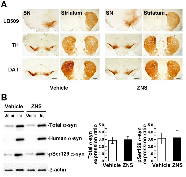
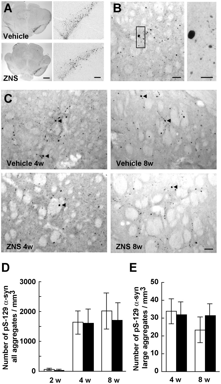
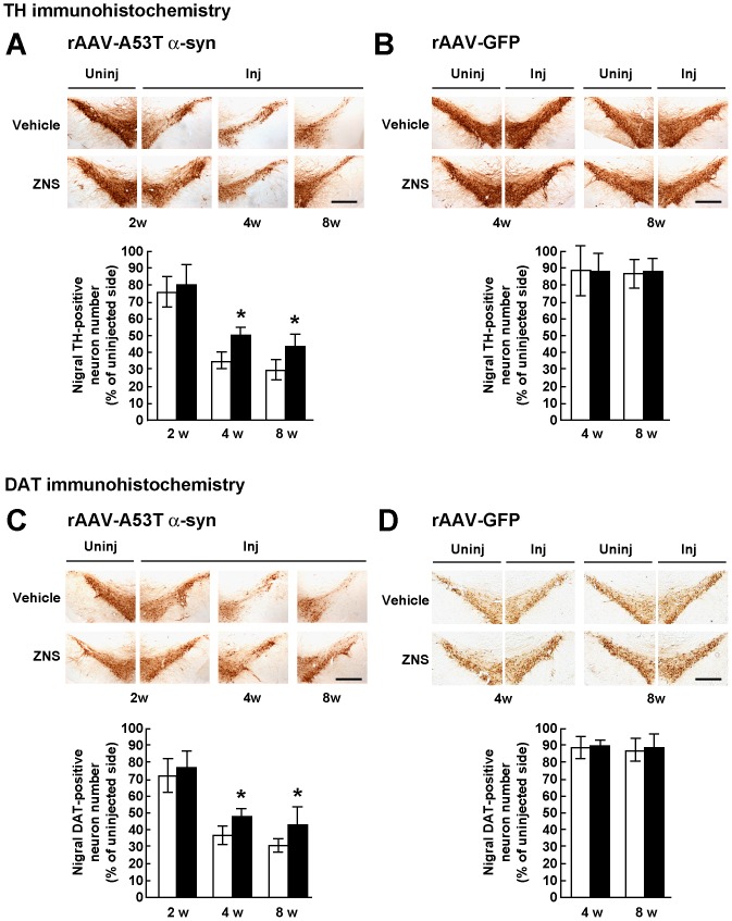
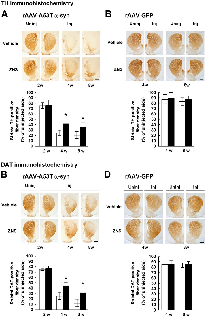
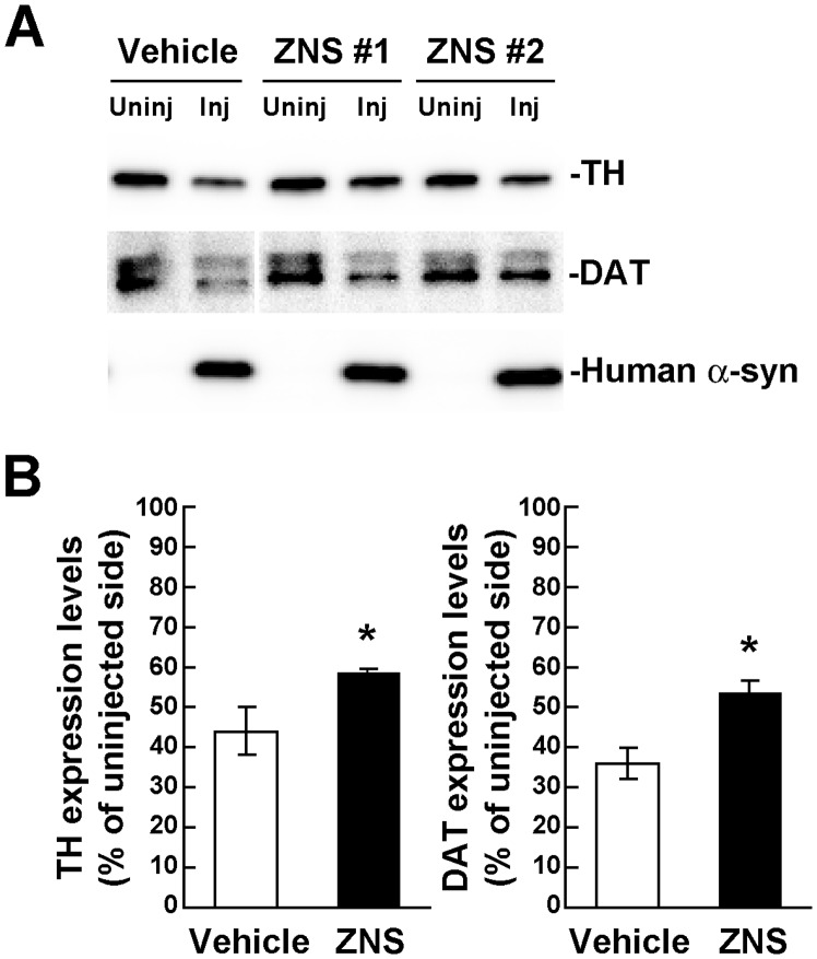
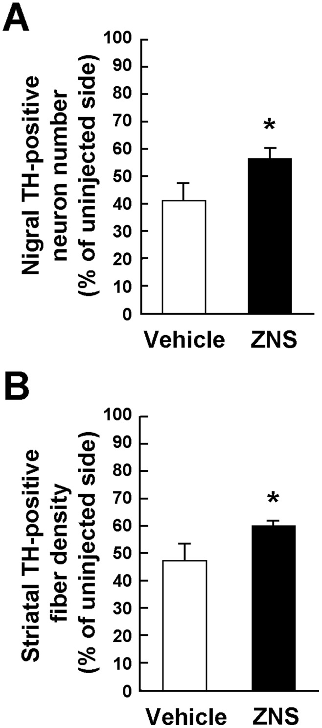
The anti-epileptic agent zonisamide (ZNS) has been shown to exert protective effects in neurotoxin-based mouse models of Parkinson disease. However, it is unknown whether ZNS can attenuate toxicity of familial Parkinson's disease-causing gene products. In this study, we investigated the effects of ZNS on neurodegeneration induced by expression of A53T α-synuclein in the rat substantia nigra using a recombinant adeno-associated virus vector. Expression of A53T α-synuclein yielded severe loss of nigral dopamine neurons and striatal dopamine nerve terminals from 2 weeks to 4 weeks after viral injection. Oral administration of ZNS (40 mg/kg/day) significantly delayed the pace of degeneration at 4 weeks after viral injection as compared with the vehicle group. This effect lasted until 8 weeks after viral injection, the final point of observation. ZNS treatment had no impact on the survival of nigrostriatal dopamine neurons in rats expressing green fluorescent protein. Quantification of striatal Ser129-phosphorylated α-synuclein-positive aggregates showed that these aggregates rapidly formed from 2 weeks to 4 weeks after viral injection. This increase was closely correlated with loss of nigrostriatal dopamine neurons. However, ZNS treatment failed to alter the number of all striatal Ser129-phosphorylated α-synuclein-positive aggregates, including small dot-like and large round structures. The number of these aggregates was almost constant at 4 weeks and 8 weeks after viral injection, although ZNS persistently prevented loss of nigrostriatal dopamine neurons during this period. Also, ZNS treatment did not affect the number of striatal aggregates larger than 10 µm in diameter. These data show that ZNS attenuates α-synuclein-induced toxicity in a manner that is independent of the formation and maturation of α-synuclein aggregates in an in vivo model of familial Parkinson's disease, suggesting that ZNS may protect nigrostriatal dopamine neurons by modulating cellular damage or a cell death pathway commonly caused by neurotoxins and α-synuclein.
抗癫痫药物唑尼沙胺(ZNS)已被证明在基于神经毒素的帕金森病小鼠模型中具有保护作用。然而,尚不清楚ZNS是否能减轻家族性帕金森病致病基因产物的毒性。在本研究中,我们使用重组腺相关病毒载体,研究了ZNS对大鼠黑质中由A53T α-突触核蛋白表达诱导的神经退行性变的影响。病毒注射后2周4周,A53T α-突触核蛋白的表达导致黑质多巴胺能神经元和纹状体多巴胺神经末梢严重丧失。与溶剂对照组相比,口服ZNS(40 mg/kg/天)显著延缓了病毒注射后4周时的退变速度。这种作用持续到病毒注射后8周,即观察的最后时间点。ZNS治疗对表达绿色荧光蛋白的大鼠黑质纹状体多巴胺能神经元的存活没有影响。对纹状体Ser129磷酸化α-突触核蛋白阳性聚集体的定量分析表明,这些聚集体在病毒注射后2周4周迅速形成。这种增加与黑质纹状体多巴胺能神经元的丧失密切相关。然而,ZNS治疗未能改变所有纹状体Ser129磷酸化α-突触核蛋白阳性聚集体的数量,包括小斑点状和大圆形结构。尽管在此期间ZNS持续阻止黑质纹状体多巴胺能神经元的丧失,但这些聚集体的数量在病毒注射后4周和8周时几乎保持不变。此外,ZNS治疗对直径大于10 µm的纹状体聚集体数量没有影响。这些数据表明,在家族性帕金森病的体内模型中,ZNS以一种独立于α-突触核蛋白聚集体形成和成熟的方式减轻α-突触核蛋白诱导的毒性,这表明ZNS可能通过调节通常由神经毒素和α-突触核蛋白引起的细胞损伤或细胞死亡途径来保护黑质纹状体多巴胺能神经元。