Zhang Song, An Qingzhu, Li Qianyun, Huang Jun, Chen Xi, Chen Xiaoyan, Zhang Jun, Wang Yongting, Yang Guo-Yuan, Zhu Wei
Department of Neurosurgery, Huashan Hospital, Fudan University, Shanghai, China.
Neuroscience and Neuroengineering Research Center, Med-X Research Institute and School of Biomedical Engineering, Shanghai Jiao Tong University, Shanghai, China.
PLoS One. 2014 Feb 28;9(2):e90069. doi: 10.1371/journal.pone.0090069. eCollection 2014.
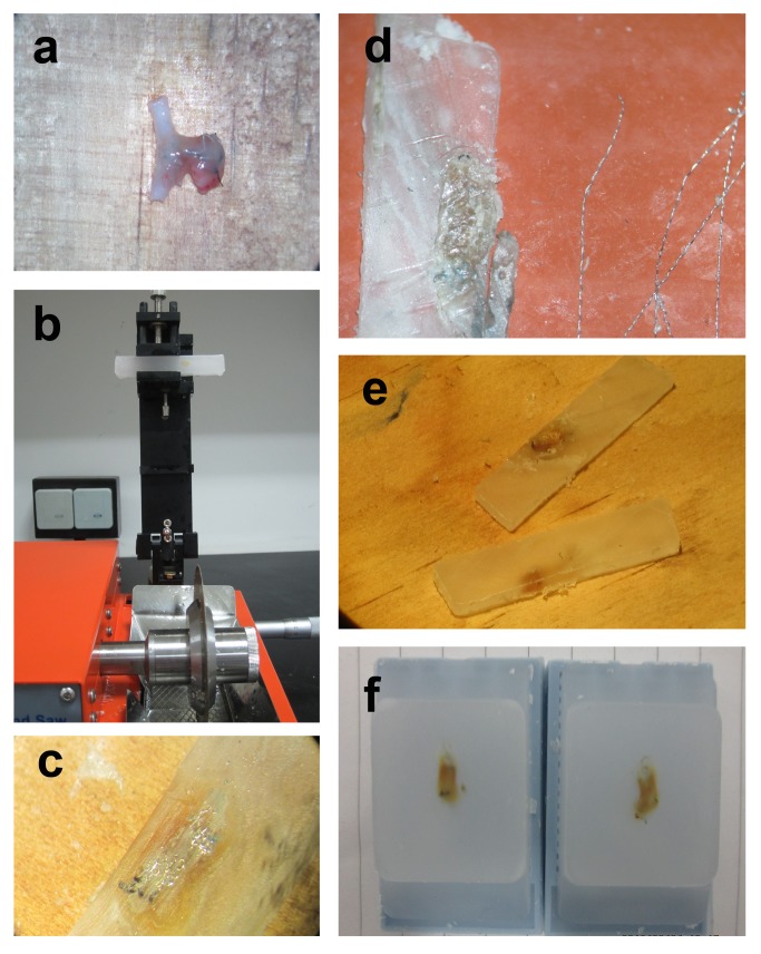
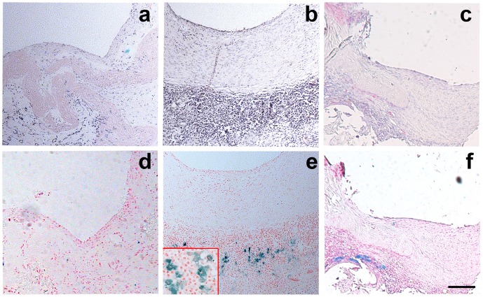
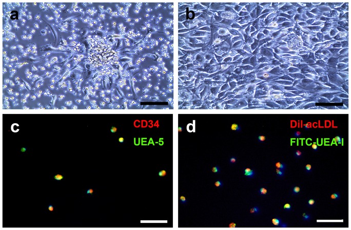
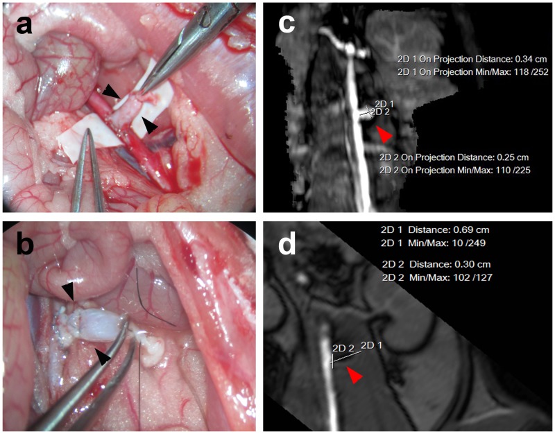
Aneurysm embolization with coil is now widely used clinically. However, the recurrence of aneurysms after embolization has always plagued neurosurgeons because the endothelial layer of the aneurysm neck loses its integrity after being embolized by coil. Bone marrow-derived endothelial progenitor cells (BM-EPCs) could be incorporated into injured endothelium and differentiate into mature endothelial cells during vascular repairing processes. The aim of our study is to explore the effects of BM-EPCs on aneurysm repairing and remodeling in a rat embolization model of abdominal aortic aneurysm. BM-EPC proliferation, migration and tube formation were not affected by super-paramagnetic iron oxide nanoparticle (SPIO) labeling compared to the controls (p>0.05). The number of SPIO-labeled cells greatly increased in EPC transplanted rats compared to that of phosphate buffered saline treated rats. SPIO-labeled EPC (SPIO-EPC) are mainly located in the aneurysm neck and surrounded by fibrous tissue. A histology study showed that the aneurysm orifice was closed with neointima and the aneurysm was filled with newly formed fibrous tissue. The SPIO-EPC accumulated in the aneurysm neck, which accelerated focal fibrous tissue remodeling, suggesting that BM-EPCs play a crucial role in repairing and remodeling the aneurysm neck orifice.
目前,弹簧圈栓塞动脉瘤在临床上已被广泛应用。然而,栓塞后动脉瘤的复发一直困扰着神经外科医生,因为动脉瘤颈部的内皮细胞层在被弹簧圈栓塞后失去了完整性。骨髓来源的内皮祖细胞(BM-EPCs)在血管修复过程中可整合到受损的内皮中,并分化为成熟的内皮细胞。本研究的目的是在大鼠腹主动脉瘤栓塞模型中探讨BM-EPCs对动脉瘤修复和重塑的影响。与对照组相比,超顺磁性氧化铁纳米颗粒(SPIO)标记对BM-EPC的增殖、迁移和管腔形成没有影响(p>0.05)。与磷酸盐缓冲盐水处理的大鼠相比,EPC移植大鼠中SPIO标记的细胞数量大大增加。SPIO标记的EPC(SPIO-EPC)主要位于动脉瘤颈部,并被纤维组织包围。组织学研究表明,动脉瘤口被新生内膜封闭,动脉瘤内充满新形成的纤维组织。SPIO-EPC聚集在动脉瘤颈部,加速了局部纤维组织重塑,提示BM-EPCs在动脉瘤颈部开口的修复和重塑中起关键作用。