Park Mee Jin, Kim So Yeon, Yoon Sang Min, Kim Jong Hoon, Park Seong Ho, Lee Seung Soo, Lee Yedaun, Lee Moon-Gyu
Department of Radiology and Research Institute of Radiology, University of Ulsan College of Medicine, Asan Medical Center, Seoul, Korea.
Department of Radiation Oncology, University of Ulsan College of Medicine, Asan Medical Center, Seoul, Korea.
PLoS One. 2014 Feb 28;9(2):e90327. doi: 10.1371/journal.pone.0090327. eCollection 2014.
To evaluate temporal changes in contrast enhancement patterns of non-tumorous hepatic parenchyma with a focus on arterial hypervascularity on multiphase computed tomography (CT) in patients with hepatocellular carcinoma (HCC) treated with stereotactic body radiotherapy (SBRT).
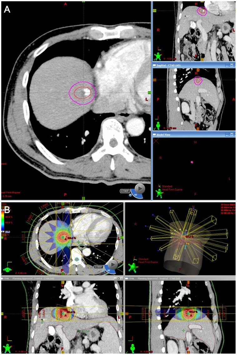
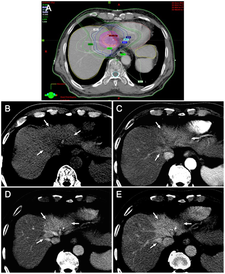
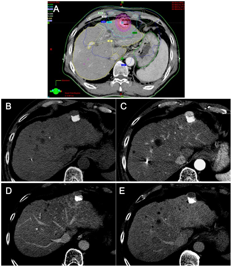
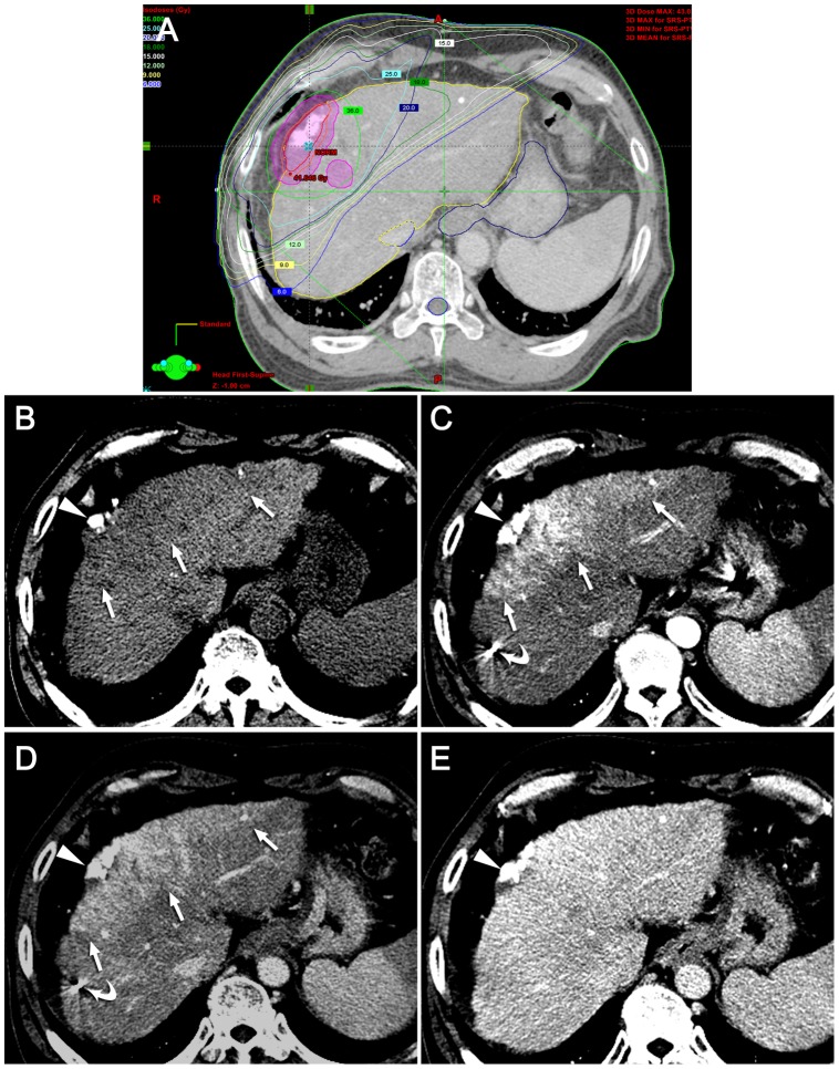
We retrospectively identified 61 patients who had undergone multiphase contrast-enhanced CT at one, three, and six months after SBRT. Irradiated versus non-irradiated liver parenchyma was delineated by cross-correlation with the dose-volume histogram of SBRT plan. Serial changes in the contrast enhancement patterns of the irradiated versus non-irradiated liver parenchyma were evaluated by two abdominal radiologists in consensus. We compared the frequency of the contrast enhancement patterns according to the follow-up period using the Fisher-Freeman-Halton exact test.
The irradiated non-tumorous hepatic parenchyma showed that the prevalence of arterial hypervascularity increased during the follow-up period (P<.01): 11.5% (7/61) in one, 45.9% (28/61) in three, and 54.1% (33/61) in six months. Contrast wash-out on the delayed phase was uncommon: 1.6% (1/61) in one, 3.3% (2/61) in three, and 0% in six months.
The incidence of arterial hypervascularity of the irradiated hepatic parenchyma gradually increased until six months after SBRT, which could interfere with the accurate evaluation of treatment response. The lack of wash-out on the delayed phase in the hypervascular area would distinguish SBRT-related change from residual/recurred HCC.
评估立体定向体部放疗(SBRT)治疗的肝细胞癌(HCC)患者非肿瘤性肝实质对比增强模式的时间变化,重点关注多期计算机断层扫描(CT)上的动脉期高灌注。
我们回顾性纳入了61例在SBRT后1个月、3个月和6个月接受多期对比增强CT检查的患者。通过与SBRT计划的剂量体积直方图进行互相关分析,勾画出受照射与未受照射的肝实质。由两名腹部放射科医生共同评估受照射与未受照射肝实质对比增强模式的系列变化。我们使用Fisher-Freeman-Halton精确检验比较随访期间对比增强模式的频率。
受照射的非肿瘤性肝实质显示,随访期间动脉期高灌注的发生率增加(P<0.01):1个月时为11.5%(7/61),3个月时为45.9%(28/61),6个月时为54.1%(33/61)。延迟期对比剂廓清不常见:1个月时为1.6%(1/61),3个月时为3.3%(2/61),6个月时为0%。
受照射肝实质的动脉期高灌注发生率在SBRT后6个月内逐渐增加,这可能会干扰对治疗反应的准确评估。高灌注区域延迟期无廓清可将SBRT相关改变与残留/复发的HCC区分开来。