Korenić Andrej, Boltze Johannes, Deten Alexander, Peters Myriam, Andjus Pavle, Radenović Lidija
Centre for Laser Microscopy, Department of Physiology and Biochemistry, Faculty of Biology, University of Belgrade, Belgrade, Serbia.
Fraunhofer Institute for Cell Therapy and Immunology, Leipzig, Germany ; Translational Centre for Regenerative Medicine, University of Leipzig, Leipzig, Germany ; Massachusetts General Hospital and Harvard Medical School, Boston, Massachusetts, United States of America.
PLoS One. 2014 Feb 28;9(2):e90697. doi: 10.1371/journal.pone.0090697. eCollection 2014.
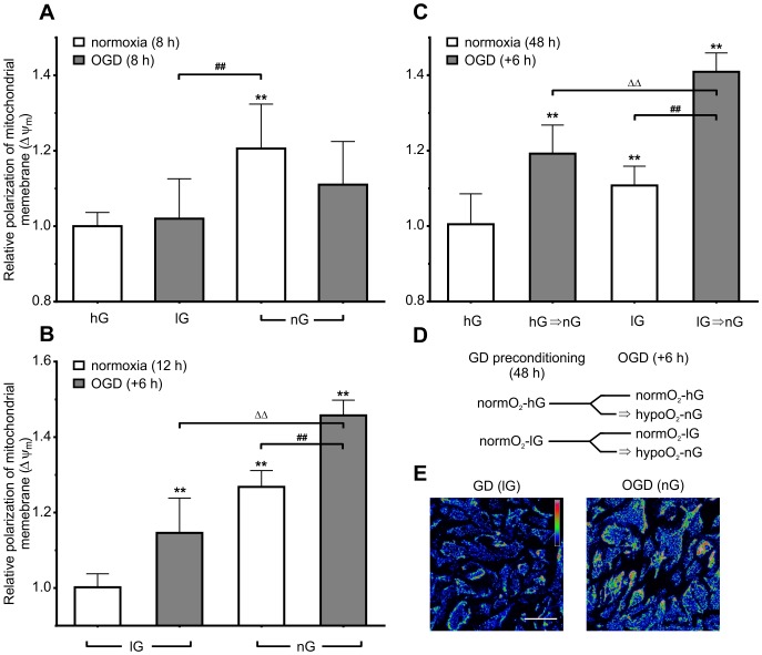
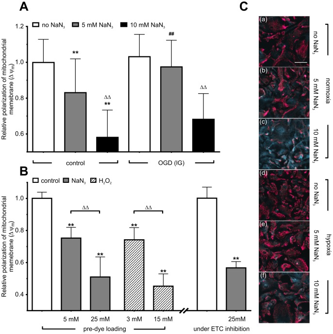
Astrocytes can tolerate longer periods of oxygen and glucose deprivation (OGD) as compared to neurons. The reasons for this reduced vulnerability are not well understood. Particularly, changes in mitochondrial membrane potential (Δψ(m)) in astrocytes, an indicator of the cellular redox state, have not been investigated during reperfusion after extended OGD exposure. Here, we subjected primary mouse astrocytes to glucose deprivation (GD), OGD and combinations of both conditions varying in duration and sequence. Changes in Δψ(m), visualized by change in the fluorescence of JC-1, were investigated within one hour after reconstitution of oxygen and glucose supply, intended to model in vivo reperfusion. In all experiments, astrocytes showed resilience to extended periods of OGD, which had little effect on Δψ(m) during reperfusion, whereas GD caused a robust Δψ(m) negativation. In case no Δψ(m) negativation was observed after OGD, subsequent chemical oxygen deprivation (OD) induced by sodium azide caused depolarization, which, however, was significantly delayed as compared to normoxic group. When GD preceded OD for 12 h, Δψ(m) hyperpolarization was induced by both GD and subsequent OD, but significant interaction between these conditions was not detected. However, when GD was extended to 48 h preceding OGD, hyperpolarization enhanced during reperfusion. This implicates synergistic effects of both conditions in that sequence. These findings provide novel information regarding the role of the two main substrates of electron transport chain (glucose and oxygen) and their hyperpolarizing effect on Δψ(m) during substrate deprivation, thus shedding new light on mechanisms of astrocyte resilience to prolonged ischemic injury.
与神经元相比,星形胶质细胞能够耐受更长时间的氧糖剥夺(OGD)。这种较低易损性的原因尚不清楚。特别是,在长时间OGD暴露后的再灌注过程中,尚未研究作为细胞氧化还原状态指标的星形胶质细胞线粒体膜电位(Δψ(m))的变化。在此,我们使原代小鼠星形胶质细胞经历葡萄糖剥夺(GD)、OGD以及这两种条件在持续时间和顺序上的不同组合。通过JC-1荧光变化可视化的Δψ(m)变化,在恢复氧气和葡萄糖供应后的一小时内进行研究,旨在模拟体内再灌注。在所有实验中,星形胶质细胞对长时间的OGD表现出耐受性,这对再灌注期间的Δψ(m)影响很小,而GD则导致Δψ(m)显著降低。如果在OGD后未观察到Δψ(m)降低,随后由叠氮化钠诱导的化学性氧剥夺(OD)会导致去极化,然而,与常氧组相比,这种去极化明显延迟。当GD在OD之前持续12小时时,GD和随后的OD均诱导Δψ(m)超极化,但未检测到这些条件之间的显著相互作用。然而,当在OGD之前将GD延长至48小时时,再灌注期间超极化增强。这表明这两种条件按该顺序具有协同作用。这些发现提供了关于电子传递链的两种主要底物(葡萄糖和氧气)的作用及其在底物剥夺期间对Δψ(m)的超极化作用的新信息,从而为星形胶质细胞对长期缺血性损伤的耐受性机制提供了新的线索。