University of California, Berkeley and University of California, San Francisco Graduate Program in Bioengineering, University of California, Berkeley/San Francisco, CA, USA; Department of Radiology and Biomedical Imaging, University of California, San Francisco (UCSF), CA, USA.
NMR Biomed. 2014 May;27(5):578-93. doi: 10.1002/nbm.3097. Epub 2014 Mar 5.
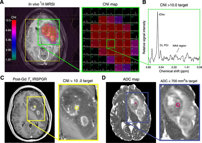
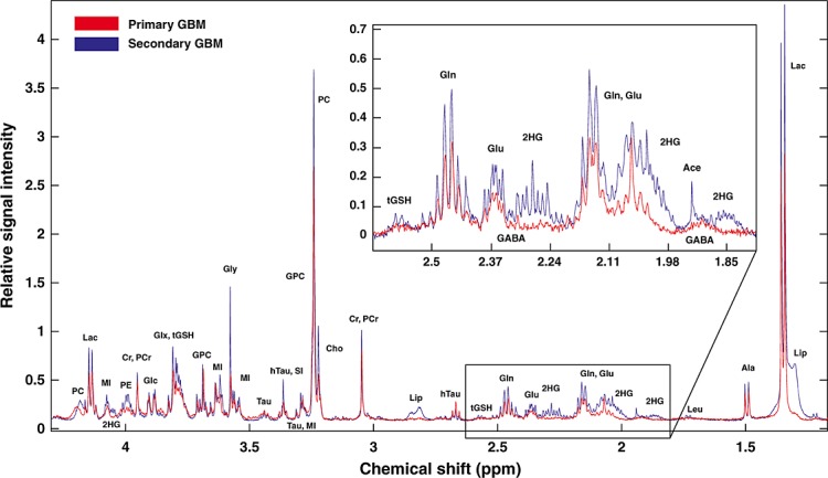
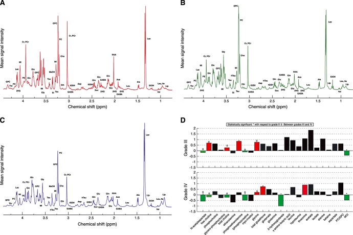
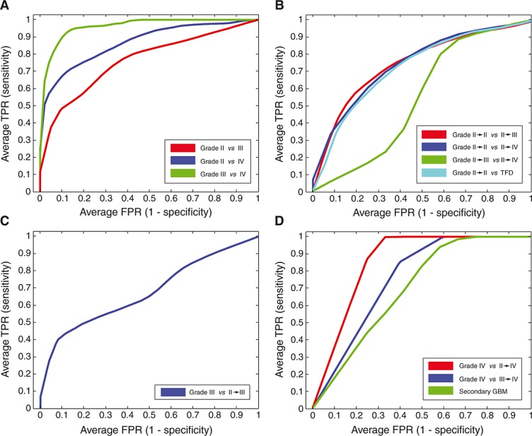
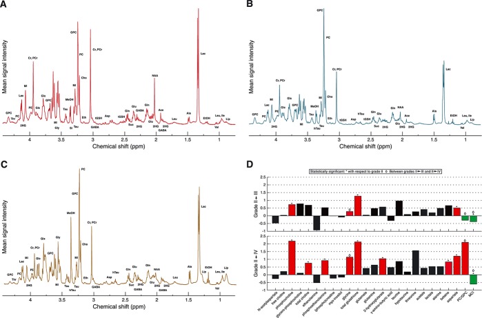
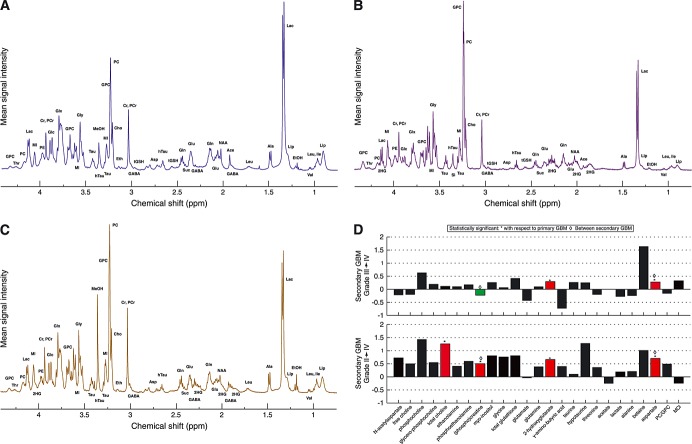
Gliomas are routinely graded according to histopathological criteria established by the World Health Organization. Although this classification can be used to understand some of the variance in the clinical outcome of patients, there is still substantial heterogeneity within and between lesions of the same grade. This study evaluated image-guided tissue samples acquired from a large cohort of patients presenting with either new or recurrent gliomas of grades II-IV using ex vivo proton high-resolution magic angle spinning spectroscopy. The quantification of metabolite levels revealed several discrete profiles associated with primary glioma subtypes, as well as secondary subtypes that had undergone transformation to a higher grade at the time of recurrence. Statistical modeling further demonstrated that these metabolomic profiles could be differentially classified with respect to pathological grading and inter-grade conversions. Importantly, the myo-inositol to total choline index allowed for a separation of recurrent low-grade gliomas on different pathological trajectories, the heightened ratio of phosphocholine to glycerophosphocholine uniformly characterized several forms of glioblastoma multiforme, and the onco-metabolite D-2-hydroxyglutarate was shown to help distinguish secondary from primary grade IV glioma, as well as grade II and III from grade IV glioma. These data provide evidence that metabolite levels are of interest in the assessment of both intra-grade and intra-lesional malignancy. Such information could be used to enhance the diagnostic specificity of in vivo spectroscopy and to aid in the selection of the most appropriate therapy for individual patients.
神经胶质瘤通常根据世界卫生组织制定的组织病理学标准进行分级。虽然这种分类可以用来理解患者临床转归的一些差异,但同一级别病变之间和内部仍然存在很大的异质性。本研究使用体外质子高分辨率魔角旋转波谱分析,评估了来自大量新发病例或复发性 II-IV 级神经胶质瘤患者的图像引导组织样本。代谢物水平的定量分析揭示了与原发性神经胶质瘤亚型相关的几个离散谱,以及在复发时已转变为更高级别(继发性)的亚型。统计模型进一步表明,这些代谢组学谱可以根据病理分级和分级间转化进行差异分类。重要的是,肌醇与总胆碱指数可区分不同病理轨迹的复发性低级别神经胶质瘤,磷酸胆碱与甘油磷酸胆碱的高比值均匀地描述了几种多形性胶质母细胞瘤的形式,而肿瘤代谢物 D-2-羟基戊二酸被证明有助于区分继发性和原发性 IV 级神经胶质瘤,以及 II 级和 III 级与 IV 级神经胶质瘤。这些数据表明代谢物水平在评估肿瘤内和肿瘤内恶性程度方面具有重要意义。这些信息可用于提高体内波谱的诊断特异性,并有助于为个体患者选择最合适的治疗方法。