Brothers Holly M, Bardou Isabelle, Hopp Sarah C, Marchalant Yannick, Kaercher Roxanne M, Turner Sarah M, Mitchem Mollie R, Kigerl Kristina, Wenk Gary L
Department of Psychology, Ohio State University, Columbus, OH, USA.
Department of Neuroscience, Ohio State University, Columbus, OH, USA.
J Alzheimers Dis Parkinsonism. 2013 Mar 28;3:110. doi: 10.4172/2161-0460.1000110.
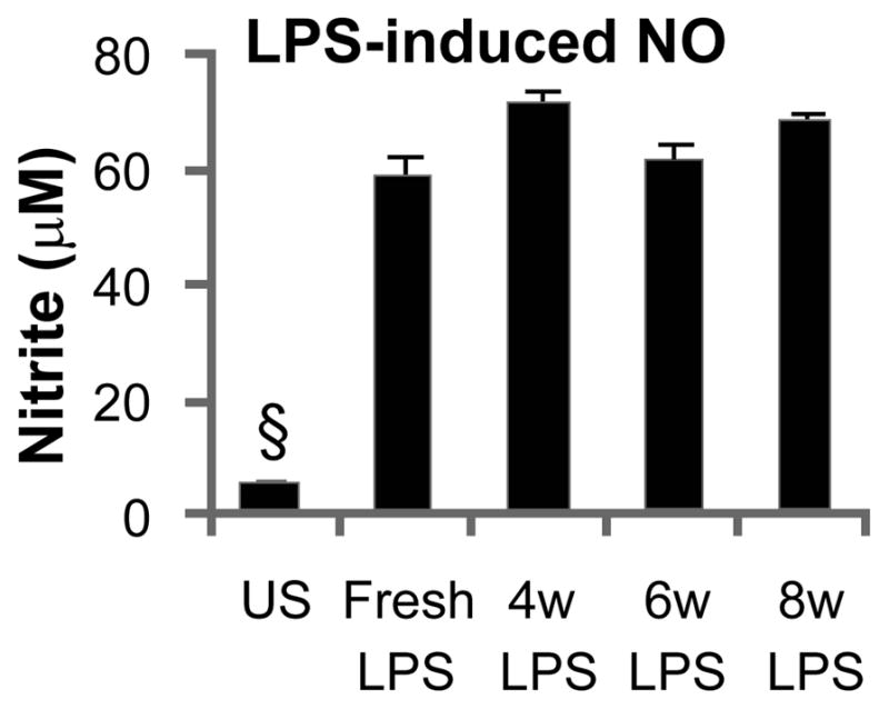
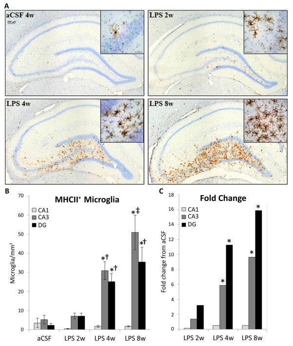
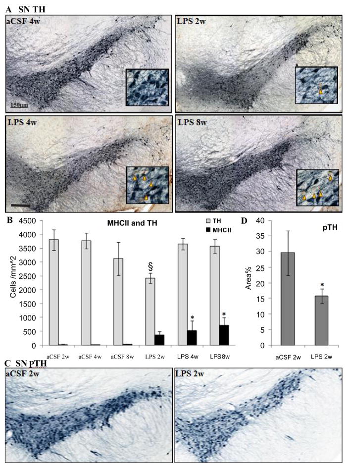
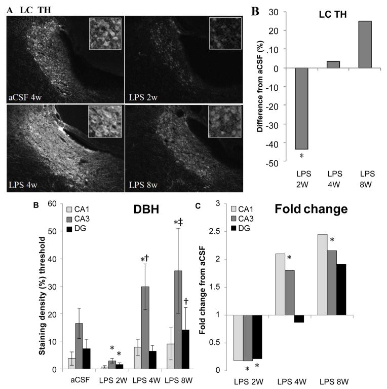
Chronic neuroinflammation is characteristic of neurodegenerative diseases and is present during very early stages, yet significant pathology and behavioral deficits do not manifest until advanced age. We investigated the consequences of experimentally-induced chronic neuroinflammation within the hippocampus and brainstem of young (4 mo) F-344 rats. Lipopolysaccharide (LPS) was infused continuously into the IV ventricle for 2, 4 or 8 weeks. The number of MHC II immunoreactive microglia in the brain continued to increase throughout the infusion period. In contrast, performance in the Morris water maze was impaired after 4 weeks but recovered by 8 weeks. Likewise, a transient loss of tyrosine hydroxylase immunoreactivity in the substantia nigra and locus coeruleus was observed after 2 weeks, but returned to control levels by 4 weeks of continuous LPS infusion. These data suggest that direct activation of microglia is sufficient to drive, but not sustain, spatial memory impairment and a decrease in tyrosine hydroxylase production in young rats. Our previous studies suggest that chronic neuroinflammation elevates extracellular glutamate and that this elevation underlies the spatial memory impairment. In the current study, increased levels of GLT1 and SNAP25 in the hippocampus corresponded with the resolution of performance deficit. Increased expression of SNAP25 is consistent with reduced glutamate release from axonal terminals while increased GLT1 is consistent with enhanced clearance of extracellular glutamate. These data demonstrate the capacity of the brain to compensate for the presence of chronic neuroinflammation, despite continued activation of microglia, through changes in the regulation of the glutamatergic system.
慢性神经炎症是神经退行性疾病的特征,且在疾病早期就已存在,但显著的病理学改变和行为缺陷直到老年才会显现。我们研究了在年轻(4个月)的F-344大鼠海马体和脑干中实验性诱导的慢性神经炎症的后果。将脂多糖(LPS)持续注入第四脑室2、4或8周。在整个注入期间,大脑中MHC II免疫反应性小胶质细胞的数量持续增加。相比之下,莫里斯水迷宫实验中的表现在4周后受损,但在8周时恢复。同样,在持续注入LPS 2周后观察到黑质和蓝斑中酪氨酸羟化酶免疫反应性短暂丧失,但在4周时恢复到对照水平。这些数据表明,小胶质细胞的直接激活足以引发但不能维持幼鼠的空间记忆损害和酪氨酸羟化酶产生的减少。我们先前研究表明,慢性神经炎症会升高细胞外谷氨酸水平,而这种升高是空间记忆损害的基础。在当前研究中,海马体中GLT1和SNAP25水平的升高与行为缺陷的缓解相对应。SNAP25表达增加与轴突终末谷氨酸释放减少一致,而GLT1增加与细胞外谷氨酸清除增强一致。这些数据证明,尽管小胶质细胞持续激活,但大脑能够通过改变谷氨酸能系统的调节来补偿慢性神经炎症的存在。