Shi Yawei, Zhang Lei, Yang Ting
Institute of Biotechnology, Key Laboratory of Chemical Biology and Molecular Engineering of Ministry of Education, Shanxi University, Taiyuan, Shanxi, P. R. China.
Institute of Biotechnology, Key Laboratory of Chemical Biology and Molecular Engineering of Ministry of Education, Shanxi University, Taiyuan, Shanxi, P. R. China; Institute of Clinical Medicine and Department of Cardiology, Renmin hospital, Hubei University of Medicine, Shiyan, Hubei, P. R. China.
PLoS One. 2014 Mar 14;9(3):e91953. doi: 10.1371/journal.pone.0091953. eCollection 2014.
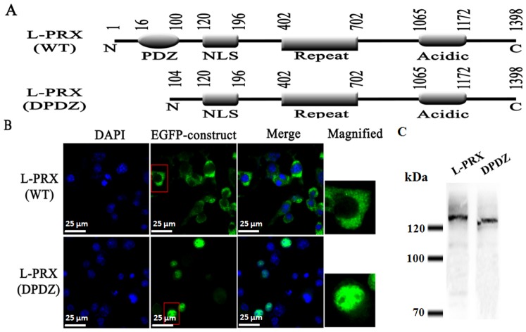
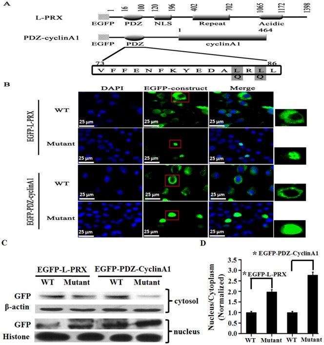
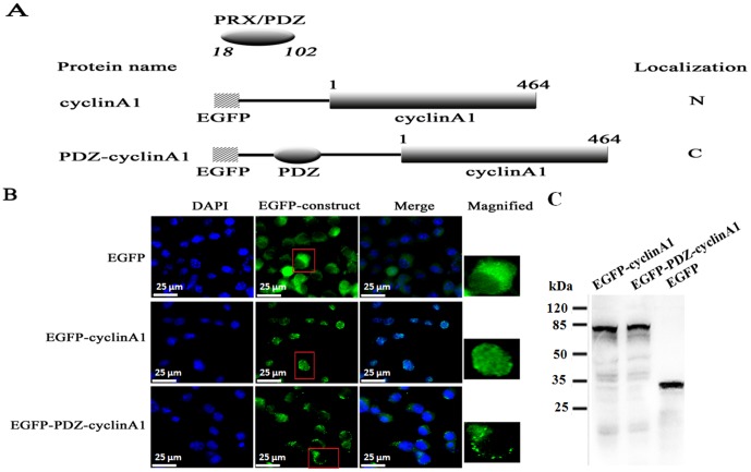
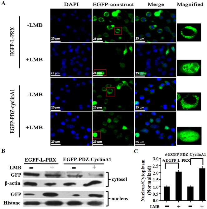
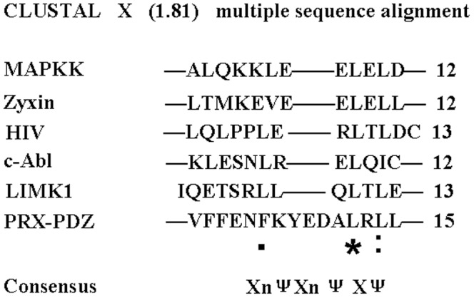
Myelinating Schwann cells specifically express L-periaxin (L-PRX) in the mammalian peripheral nervous system. Several loss-of-function mutations in periaxin have been described and linked to autosomal recessive Dejerine Sottas neuropathy and to demyelinating Charcot-Marie-Tooth disease. The localization of L-periaxin is developmentally regulated in the nucleus and the plasma membrane of Schwann cells. In this study, L-periaxin, which contains a PDZ domain, a nuclear localization signal (NLS) domain, a repeat domain, and an acidic domain, was localized in the cytoplasm of RSC96 cells. By contrast, a mutant L-periaxin with a deleted PDZ domain was localized mainly in the nucleus of RSC96 cells. After a nuclear cyclin A1, which is localized exclusively in the nucleus, was fused with the PDZ domain, cyclinA1was found in the cytoplasm of RSC96 cells. Treatment with leptomycin B (LMB), a specific inhibitor of nuclear export mediated by leucine-rich nuclear export signal (NES), also causes nuclear accumulation of wild-type L-periaxin. Double leucine mutation (L83, 85Q) in the putative NES in the PDZ domain prevented L-periaxin nuclear export and induced nuclear accumulation. These results suggested that the localization of L-periaxin in the cytoplasm is supported by NES in the PDZ domain.
在哺乳动物外周神经系统中,形成髓鞘的施万细胞特异性表达L-外周蛋白(L-PRX)。外周蛋白中的几个功能丧失突变已被描述,并与常染色体隐性德热里纳 - 索塔斯神经病和脱髓鞘型夏科 - 马里 - 图斯病相关。L-外周蛋白的定位在施万细胞的细胞核和质膜中受到发育调控。在本研究中,含有PDZ结构域、核定位信号(NLS)结构域、重复结构域和酸性结构域的L-外周蛋白定位于RSC96细胞的细胞质中。相比之下,缺失PDZ结构域的突变型L-外周蛋白主要定位于RSC96细胞的细胞核中。将仅定位于细胞核的核细胞周期蛋白A1与PDZ结构域融合后,细胞周期蛋白A1出现在RSC96细胞的细胞质中。用富含亮氨酸的核输出信号(NES)介导的核输出特异性抑制剂雷帕霉素B(LMB)处理,也会导致野生型L-外周蛋白在细胞核中积累。PDZ结构域中假定的NES中的双亮氨酸突变(L83,85Q)阻止了L-外周蛋白的核输出并诱导了核积累。这些结果表明,PDZ结构域中的NES支持L-外周蛋白在细胞质中的定位。