Aghanejad Ayuob, Jalilian Amir R, Fazaeli Yousef, Alirezapoor Behrouz, Pouladi Mehraban, Beiki Davoud, Maus Stephan, Khalaj Ali
Research Center for Nuclear Medicine, Tehran University of Medical Sciences, Tehran, Iran. ; Department of Nuclear Pharmacy, Faculty of Pharmacy, Tehran University of Medical Sciences, Tehran, Iran.
Radiation Application Research School, Nuclear Science and Technology Research Institute, Tehran, 11365-3486, Iran.
Sci Pharm. 2013 Sep 12;82(1):29-42. doi: 10.3797/scipharm.1305-18. Print 2014 Jan-Mar.
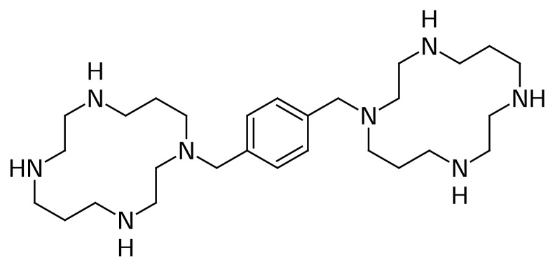
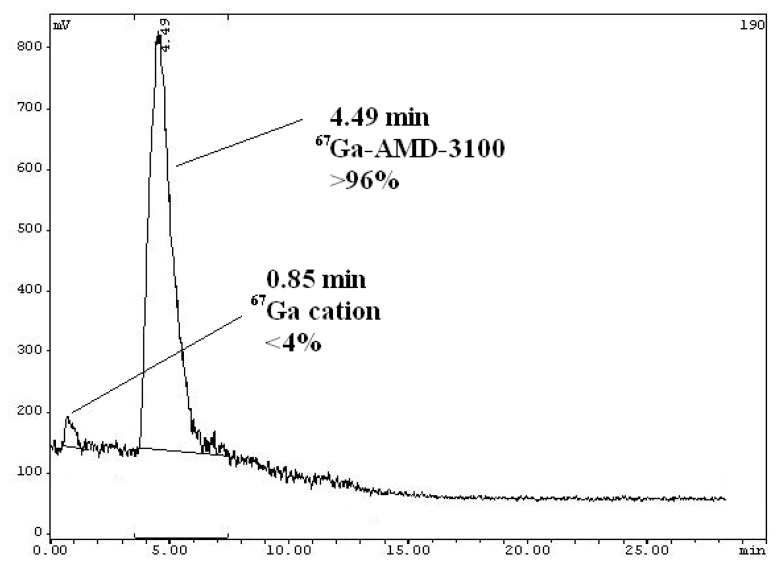
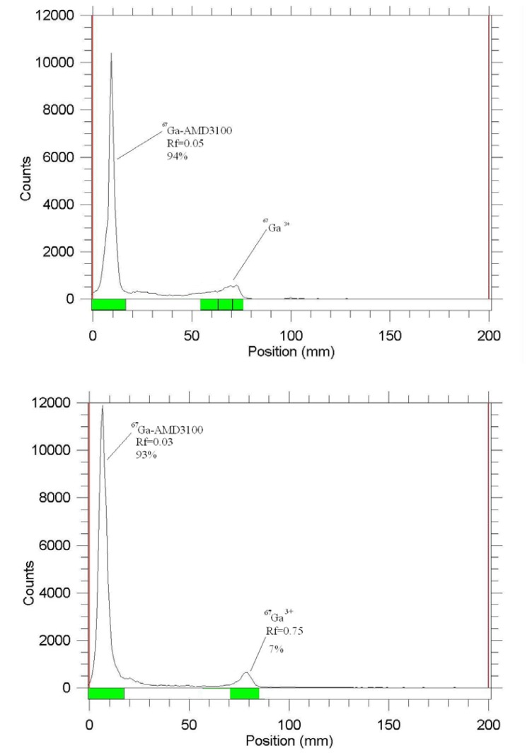
In order to develop a possible C-X-C chemokine receptor type 4 (CXCR4) imaging agent for oncological scintigraphy, [(67)Ga]-labeled 1,1'-[1,4-Phenylene-bis(methylene)]bis(1,4,8,11-tetraazacyclotetradecane) ([(67)Ga]-AMD3100) was prepared by using [(67)Ga]GaCl3 and AMD-3100 for 2 h at 50 °C (radiochemical purity: >95% ITLC, >99% HPLC, specific activity: 1800-2000 TBq/mmol) in acetate buffer. The stability of the complex was checked in the presence of human serum (37 °C) and in the final formulation for four days. The biodistribution of the labeled compound in the vital organs of wild type Sprague-Dawley rats was determined and compared with that of the free Ga(3+) cation up to 48 h. Considering the spleen as the target organ, the best target:non target ratios were obtained 48 h post-injection (spleen:blood ratio; 14.5 and spleen:muscle ratio; 88.4). Initial SPECT images and biodistribution results in the wild type rats matched each other and demonstrated rapid washout of the tracer from the urinary tract. SPECT images in human breast carcinoma-bearing mice demonstrated a detectable tumor uptake in 48 h post-injection.
为开发一种可能用于肿瘤闪烁显像的C-X-C趋化因子受体4(CXCR4)显像剂,通过在50℃下使用[(67)Ga]GaCl3和AMD-3100在醋酸盐缓冲液中反应2小时制备了[(67)Ga]标记的1,1'-[1,4-亚苯基-双(亚甲基)]双(1,4,8,11-四氮杂环十四烷)([(67)Ga]-AMD3100)(放射化学纯度:ITLC法>95%,HPLC法>99%,比活度:1800 - 2000 TBq/mmol)。在人血清存在下(37℃)以及在最终制剂中检查该配合物的稳定性,为期四天。测定标记化合物在野生型Sprague-Dawley大鼠重要器官中的生物分布,并与游离Ga(3+)阳离子的生物分布进行比较,长达48小时。以脾脏作为靶器官,注射后48小时获得了最佳的靶器官与非靶器官比值(脾脏:血液比值为14.5,脾脏:肌肉比值为88.4)。野生型大鼠的初始SPECT图像和生物分布结果相互匹配,并显示示踪剂从尿路快速清除。荷人乳腺癌小鼠的SPECT图像显示注射后48小时肿瘤有可检测到的摄取。