• 文献检索
  • 文档翻译
  • 深度研究
  • 学术资讯
  • Suppr Zotero 插件Zotero 插件
  • 邀请有礼
  • 套餐&价格
  • 历史记录
应用&插件
Suppr Zotero 插件Zotero 插件浏览器插件Mac 客户端Windows 客户端微信小程序
定价
高级版会员购买积分包购买API积分包
服务
文献检索文档翻译深度研究API 文档MCP 服务
关于我们
关于 Suppr公司介绍联系我们用户协议隐私条款
关注我们

Suppr 超能文献

核心技术专利:CN118964589B侵权必究
粤ICP备2023148730 号-1Suppr @ 2026

文献检索

告别复杂PubMed语法,用中文像聊天一样搜索,搜遍4000万医学文献。AI智能推荐,让科研检索更轻松。

立即免费搜索

文件翻译

保留排版,准确专业,支持PDF/Word/PPT等文件格式,支持 12+语言互译。

免费翻译文档

深度研究

AI帮你快速写综述,25分钟生成高质量综述,智能提取关键信息,辅助科研写作。

立即免费体验

头颈部鳞状细胞癌的影像学检查:PET/MRI 的潜在作用。

Imaging in head and neck squamous cell carcinoma: the potential role of PET/MRI.

机构信息

Department of Imaging, Division of Radiology, Geneva University Hospital, Geneva, Switzerland.

出版信息

Br J Radiol. 2014 Apr;87(1036):20130677. doi: 10.1259/bjr.20130677. Epub 2014 Feb 3.

DOI:10.1259/bjr.20130677
PMID:24649835
原文链接:https://pmc.ncbi.nlm.nih.gov/articles/PMC4067029/
Abstract

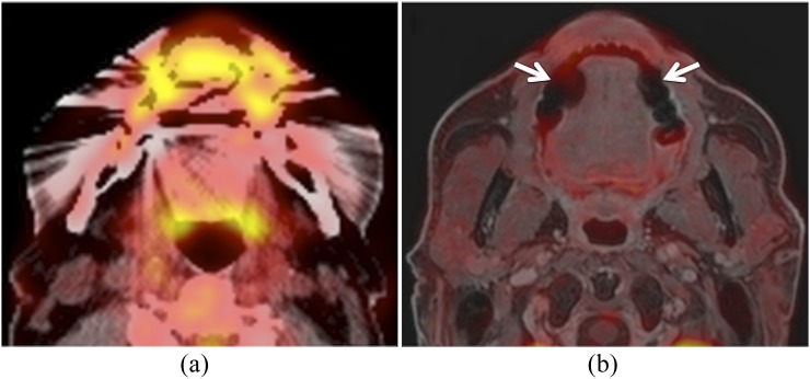
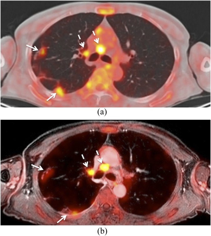
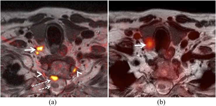
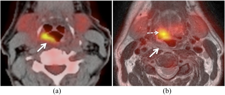
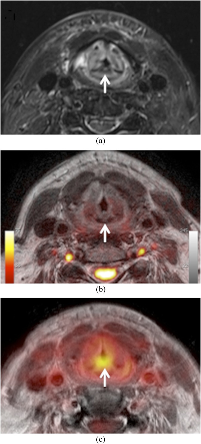
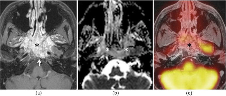
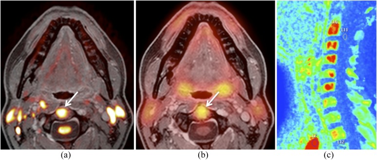
In head and neck oncology, the information provided by positron emission tomography (PET)/CT and MRI is often complementary because both the methods are based on different biophysical foundations. Therefore, combining diagnostic information from both modalities can provide additional diagnostic gain. Debates about integrated PET/MRI systems have become fashionable during the past few years, since the introduction and wide adoption of software-based multimodality image registration and fusion and the hardware implementation of integrated hybrid PET/MRI systems in pre-clinical and clinical settings. However, combining PET with MRI has proven to be technically and clinically more challenging than initially expected and, as such, research into the potential clinical role of PET/MRI in comparison with PET/CT, diffusion-weighted MRI (DW MRI) or the combination thereof is still ongoing. This review focuses on the clinical applications of PET/MRI in head and neck squamous cell carcinoma (HNSCC). We first discuss current evidence about the use of combined PET/CT and DW MRI, and, then, we explain the rationale and principles of PET/MR image fusion before summarizing the state-of-the-art knowledge regarding the diagnostic performance of PET/MRI in HNSCC. Feasibility and quantification issues, diagnostic pitfalls and challenges in clinical settings as well as ongoing research and potential future applications are also discussed.

摘要

在头颈部肿瘤学中,正电子发射断层扫描(PET)/CT 和 MRI 提供的信息通常是互补的,因为这两种方法基于不同的生物物理基础。因此,结合两种模态的诊断信息可以提供额外的诊断收益。近年来,随着基于软件的多模态图像配准和融合的引入以及在临床前和临床环境中集成混合 PET/MRI 系统的硬件实现,关于集成 PET/MRI 系统的争论变得很流行。然而,将 PET 与 MRI 结合证明比最初预期的在技术和临床方面更具挑战性,因此,与 PET/CT、扩散加权 MRI(DW MRI)或两者结合相比,研究 PET/MRI 的潜在临床作用仍在进行中。这篇综述重点介绍了 PET/MRI 在头颈部鳞状细胞癌(HNSCC)中的临床应用。我们首先讨论了关于联合使用 PET/CT 和 DW MRI 的当前证据,然后解释了 PET/MR 图像融合的原理和原则,最后总结了关于 PET/MRI 在 HNSCC 中的诊断性能的最新知识。还讨论了可行性和量化问题、诊断陷阱和临床环境中的挑战以及正在进行的研究和潜在的未来应用。

https://cdn.ncbi.nlm.nih.gov/pmc/blobs/3cec/4067029/b1844b938d32/bjr.20130677.g007.jpg
https://cdn.ncbi.nlm.nih.gov/pmc/blobs/3cec/4067029/cb343719acc9/bjr.20130677.g001.jpg
https://cdn.ncbi.nlm.nih.gov/pmc/blobs/3cec/4067029/6f4a835f3d7d/bjr.20130677.g002.jpg
https://cdn.ncbi.nlm.nih.gov/pmc/blobs/3cec/4067029/638d8f2da18f/bjr.20130677.g003.jpg
https://cdn.ncbi.nlm.nih.gov/pmc/blobs/3cec/4067029/0ea579f405b7/bjr.20130677.g004.jpg
https://cdn.ncbi.nlm.nih.gov/pmc/blobs/3cec/4067029/3010f084861c/bjr.20130677.g005.jpg
https://cdn.ncbi.nlm.nih.gov/pmc/blobs/3cec/4067029/1e492458af9e/bjr.20130677.g006.jpg
https://cdn.ncbi.nlm.nih.gov/pmc/blobs/3cec/4067029/b1844b938d32/bjr.20130677.g007.jpg

相似文献

1
Imaging in head and neck squamous cell carcinoma: the potential role of PET/MRI.头颈部鳞状细胞癌的影像学检查:PET/MRI 的潜在作用。
Br J Radiol. 2014 Apr;87(1036):20130677. doi: 10.1259/bjr.20130677. Epub 2014 Feb 3.
2
Whole-body-MR imaging including DWIBS in the work-up of patients with head and neck squamous cell carcinoma: a feasibility study.全身磁共振成像包括弥散加权体部成像在头颈部鳞状细胞癌患者检查中的应用:一项可行性研究
Eur J Radiol. 2014 Jul;83(7):1144-1151. doi: 10.1016/j.ejrad.2014.03.019. Epub 2014 Mar 30.
3
Locoregional tumour evaluation of squamous cell carcinoma in the head and neck area: a comparison between MRI, PET/CT and integrated PET/MRI.头颈部鳞状细胞癌的局部区域肿瘤评估:MRI、PET/CT与PET/MRI一体化成像的比较
Eur J Nucl Med Mol Imaging. 2016 Jan;43(1):92-102. doi: 10.1007/s00259-015-3145-z. Epub 2015 Aug 6.
4
18F-fluoro-deoxy-glucose-positron emission tomography/computed tomography in diagnosis of head and neck squamous cell carcinoma: a systematic review and meta-analysis.18F-氟脱氧葡萄糖正电子发射断层扫描/计算机断层扫描在头颈部鳞状细胞癌诊断中的应用:一项系统评价和荟萃分析
Eur J Cancer. 2014 Sep;50(13):2271-9. doi: 10.1016/j.ejca.2014.05.015. Epub 2014 Jul 7.
5
Diffusion-weighted and PET/MR Imaging after Radiation Therapy for Malignant Head and Neck Tumors.恶性头颈部肿瘤放疗后的扩散加权成像及PET/MR成像
Radiographics. 2015 Sep-Oct;35(5):1502-27. doi: 10.1148/rg.2015140029. Epub 2015 Aug 7.
6
Feasibility of Multiparametric Imaging with PET/MR in Head and Neck Squamous Cell Carcinoma.PET/MR多参数成像在头颈部鳞状细胞癌中的可行性
J Nucl Med. 2017 Jan;58(1):69-74. doi: 10.2967/jnumed.116.180091. Epub 2016 Sep 8.
7
Local recurrence of squamous cell carcinoma of the head and neck after radio(chemo)therapy: Diagnostic performance of FDG-PET/MRI with diffusion-weighted sequences.头颈部鳞状细胞癌放(化)疗后局部复发:FDG-PET/MRI 联合弥散加权序列的诊断性能。
Eur Radiol. 2018 Feb;28(2):651-663. doi: 10.1007/s00330-017-4999-1. Epub 2017 Aug 15.
8
¹⁸F-FDG PET/CT and MRI in the follow-up of head and neck squamous cell carcinoma.¹⁸F-FDG PET/CT 和 MRI 在头颈部鳞状细胞癌随访中的应用。
Contrast Media Mol Imaging. 2011 Jul-Aug;6(4):260-6. doi: 10.1002/cmmi.425.
9
Application of integrated positron emission tomography/magnetic resonance imaging in evaluating the prognostic factors of head and neck squamous cell carcinoma with positron emission tomography, diffusion-weighted imaging, dynamic contrast enhancement and combined model.正电子发射断层成像/磁共振成像在头颈部鳞癌预后因素评估中的应用——正电子发射断层成像、弥散加权成像、动态对比增强及联合模型。
Dentomaxillofac Radiol. 2020 Jul;49(5):20190488. doi: 10.1259/dmfr.20190488. Epub 2020 Apr 3.
10
Squamous Cell Carcinoma: PET/CT and PET/MRI of the Pretreatment and Post-Treatment Neck.鳞状细胞癌:治疗前和治疗后颈部的PET/CT与PET/MRI
Semin Ultrasound CT MR. 2019 Oct;40(5):400-413. doi: 10.1053/j.sult.2019.07.004. Epub 2019 Jul 17.

引用本文的文献

1
Recent advances in PET/MR imaging for head and neck tumors: a systematic review of the last three years.正电子发射断层显像/磁共振成像在头颈部肿瘤中的最新进展:过去三年的系统综述
Radiol Med. 2025 Aug 28. doi: 10.1007/s11547-025-02075-y.
2
MRI Detection of Unknown Primary Tumours in the Head and Neck: What Is the Expected Normal Asymmetry in the Size of the Palatine Tonsils?头颈部未知原发肿瘤的MRI检测:腭扁桃体大小的预期正常不对称性是多少?
Diagnostics (Basel). 2025 Mar 20;15(6):788. doi: 10.3390/diagnostics15060788.
3
Positron Emission Tomography-Magnetic Resonance Imaging, a New Hybrid Imaging Modality for Dentomaxillofacial Malignancies-A Systematic Review.

本文引用的文献

1
Utility of MRI diffusion techniques in the evaluation of tumors of the head and neck.磁共振扩散技术在头颈部肿瘤评估中的应用。
Cancers (Basel). 2013 Jul 5;5(3):875-89. doi: 10.3390/cancers5030875.
2
A study on the artifacts generated by dental materials in PET/CT image.一项关于牙科材料在PET/CT图像中产生伪影的研究。
Annu Int Conf IEEE Eng Med Biol Soc. 2013;2013:2465-8. doi: 10.1109/EMBC.2013.6610039.
3
Detection and quantification of focal uptake in head and neck tumours: (18)F-FDG PET/MR versus PET/CT.头颈部肿瘤局灶摄取的检测与定量:(18)F-FDG PET/MR 与 PET/CT 的比较。
正电子发射断层扫描-磁共振成像,一种用于牙颌面恶性肿瘤的新型混合成像模式——系统评价
Diagnostics (Basel). 2025 Mar 7;15(6):654. doi: 10.3390/diagnostics15060654.
4
Utilizing Narrow Band Imaging for Early Detection of Recurrence in Follow-Up Patients with Head and Neck Squamous Cell Carcinoma Treated with Curative Intent Using Radiotherapy/Chemo-Radiotherapy: Enhancing Detection and Patient Outcomes.利用窄带成像技术对接受根治性放疗/放化疗的头颈部鳞状细胞癌随访患者进行复发的早期检测:提高检测率及改善患者预后。
Indian J Otolaryngol Head Neck Surg. 2025 Feb;77(2):869-876. doi: 10.1007/s12070-024-05277-4. Epub 2025 Jan 3.
5
Overcoming Resistance to Standard-of-Care Therapies for Head and Neck Squamous Cell Carcinomas.克服头颈部鳞状细胞癌标准治疗的抵抗。
Cells. 2024 Jun 11;13(12):1018. doi: 10.3390/cells13121018.
6
Multiparametric Magnetic Resonance Imaging of Penile Cancer: A Pictorial Review.阴茎癌的多参数磁共振成像:图文综述
Cancers (Basel). 2023 Nov 8;15(22):5324. doi: 10.3390/cancers15225324.
7
The Prediction of Biological Features Using Magnetic Resonance Imaging in Head and Neck Squamous Cell Carcinoma: A Systematic Review and Meta-Analysis.利用磁共振成像预测头颈部鳞状细胞癌生物学特征的系统评价与Meta分析
Cancers (Basel). 2023 Oct 20;15(20):5077. doi: 10.3390/cancers15205077.
8
PET/MR versus PET/CT for locoregional staging of oropharyngeal squamous cell cancer.正电子发射断层扫描/磁共振成像(PET/MR)与正电子发射断层扫描/计算机断层扫描(PET/CT)在口咽鳞状细胞癌局部区域分期中的比较。
Acta Radiol. 2023 May;64(5):1865-1872. doi: 10.1177/02841851221140668. Epub 2022 Dec 4.
9
Diagnostic Accuracy of F-FDG-PET/CT and F-FDG-PET/MRI in Detecting Locoregional Recurrence of HNSCC 12 Weeks after the End of Chemoradiotherapy: Single-Center Experience with PET/MRI.氟代脱氧葡萄糖正电子发射断层扫描/计算机断层扫描和氟代脱氧葡萄糖正电子发射断层扫描/磁共振成像在放化疗结束后 12 周检测头颈部鳞状细胞癌局部区域复发的诊断准确性:PET/MRI 的单中心经验。
Contrast Media Mol Imaging. 2022 Aug 24;2022:8676787. doi: 10.1155/2022/8676787. eCollection 2022.
10
Detection of distant metastases and distant second primary cancers in head and neck squamous cell carcinoma: comparison of [F]FDG PET/MRI and [F]FDG PET/CT.头颈部鳞状细胞癌远处转移及远处第二原发癌的检测:[F]FDG PET/MRI与[F]FDG PET/CT的比较
Insights Imaging. 2022 Jul 28;13(1):121. doi: 10.1186/s13244-022-01261-0.
Eur J Nucl Med Mol Imaging. 2014 Mar;41(3):462-75. doi: 10.1007/s00259-013-2580-y. Epub 2013 Oct 10.
4
Pathology-based validation of FDG PET segmentation tools for volume assessment of lymph node metastases from head and neck cancer.基于病理的 FDG PET 分割工具对头颈部癌淋巴结转移体积评估的验证。
Eur J Nucl Med Mol Imaging. 2013 Dec;40(12):1828-35. doi: 10.1007/s00259-013-2513-9. Epub 2013 Aug 14.
5
Value of retrospective image fusion of ¹⁸F-FDG PET and MRI for preoperative staging of head and neck cancer: comparison with PET/CT and contrast-enhanced neck MRI.¹⁸F-FDG PET 与 MRI 回顾性图像融合对头颈部癌术前分期的价值:与 PET/CT 和增强型颈部 MRI 的比较。
Eur J Radiol. 2013 Nov;82(11):2005-10. doi: 10.1016/j.ejrad.2013.06.025. Epub 2013 Jul 24.
6
A strategy for multimodal deformable image registration to integrate PET/MR into radiotherapy treatment planning.一种将 PET/MR 整合到放射治疗计划中的多模态可变形图像配准策略。
Acta Oncol. 2013 Oct;52(7):1353-9. doi: 10.3109/0284186X.2013.813964. Epub 2013 Jul 23.
7
PET/CT versus body coil PET/MRI: how low can you go?PET/CT 与体线圈 PET/MRI:能低到何种程度?
Insights Imaging. 2013 Aug;4(4):481-90. doi: 10.1007/s13244-013-0247-7. Epub 2013 May 15.
8
Positron emission tomography (PET) attenuation correction artefacts in PET/CT and PET/MRI.正电子发射断层扫描(PET)衰减校正伪影在 PET/CT 和 PET/MRI 中的应用。
Br J Radiol. 2013 May;86(1025):20120570. doi: 10.1259/bjr.20120570.
9
MR-driven metal artifact reduction in PET/CT.MR 驱动的 PET/CT 中的金属伪影降低。
Phys Med Biol. 2013 Apr 7;58(7):2267-80. doi: 10.1088/0031-9155/58/7/2267. Epub 2013 Mar 12.
10
Functional imaging of head and neck squamous cell carcinoma with diffusion-weighted MRI and FDG PET/CT: quantitative analysis of ADC and SUV.头颈部鳞状细胞癌的扩散加权 MRI 和 FDG PET/CT 功能成像:ADC 和 SUV 的定量分析。
Eur J Nucl Med Mol Imaging. 2013 Jun;40(6):842-52. doi: 10.1007/s00259-013-2351-9. Epub 2013 Feb 22.