Callaghan Martina F, Freund Patrick, Draganski Bogdan, Anderson Elaine, Cappelletti Marinella, Chowdhury Rumana, Diedrichsen Joern, Fitzgerald Thomas H B, Smittenaar Peter, Helms Gunther, Lutti Antoine, Weiskopf Nikolaus
Wellcome Trust Centre for Neuroimaging, UCL Institute of Neurology, London, UK.
Wellcome Trust Centre for Neuroimaging, UCL Institute of Neurology, London, UK; Spinal Cord Injury Center Balgrist, University Hospital Zurich, Zurich, Switzerland; Department of Brain Repair and Rehabilitation, UCL Institute of Neurology, London, UK.
Neurobiol Aging. 2014 Aug;35(8):1862-72. doi: 10.1016/j.neurobiolaging.2014.02.008. Epub 2014 Feb 15.
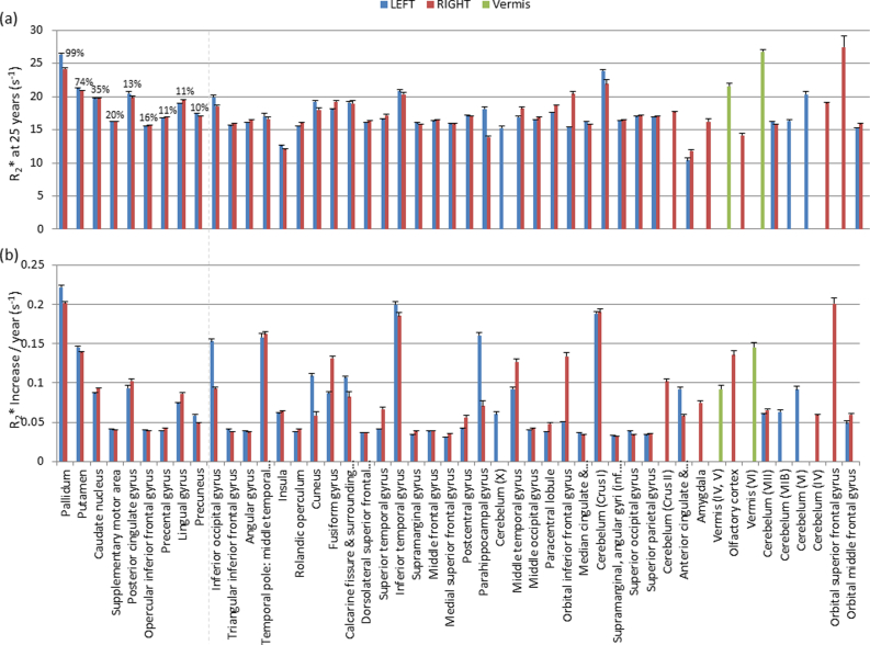
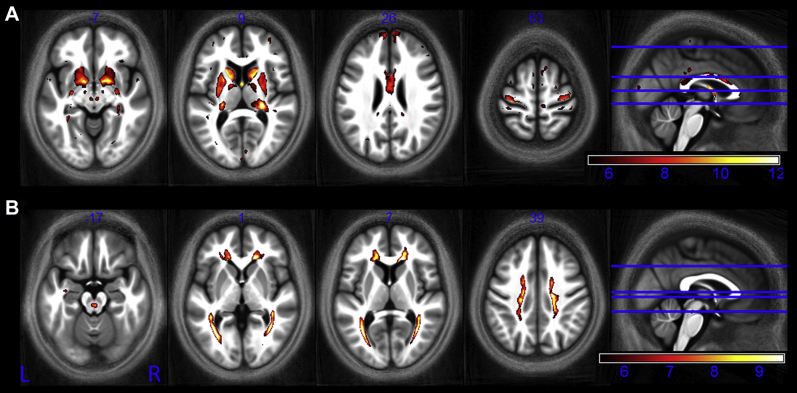
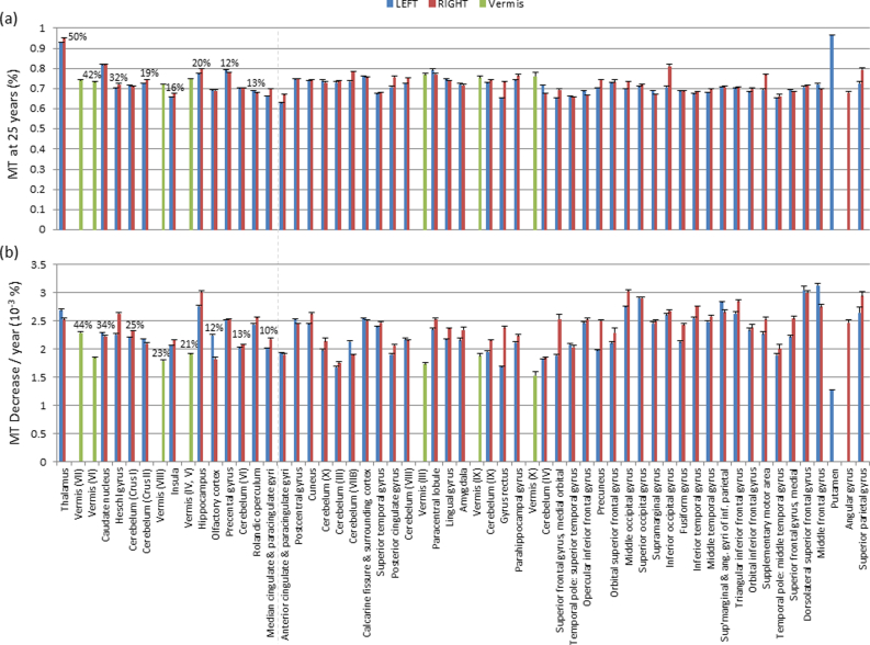
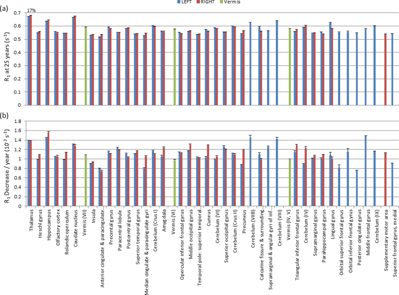
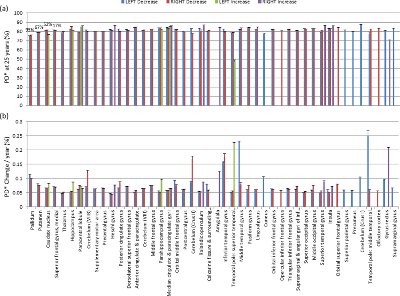
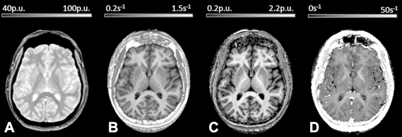
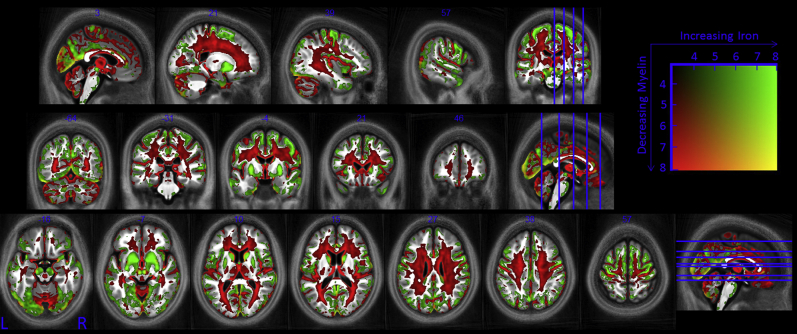
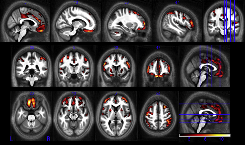
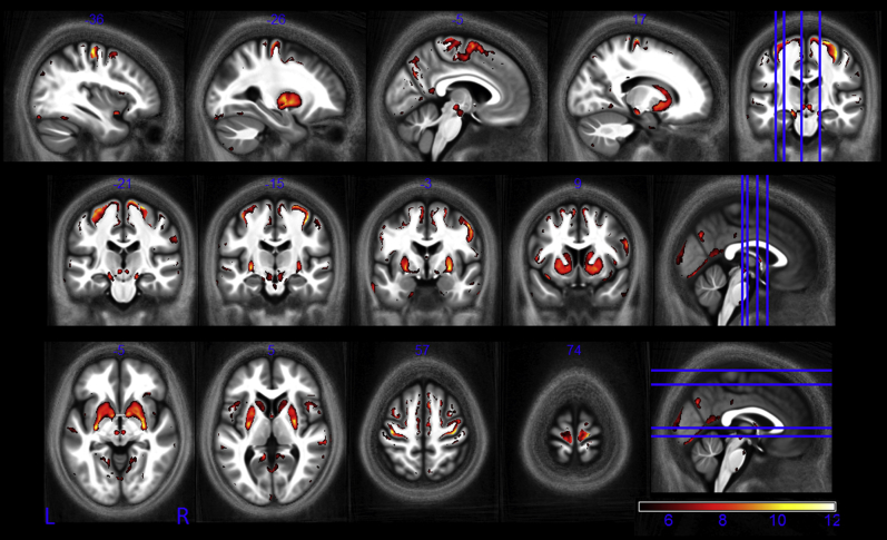
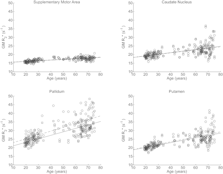
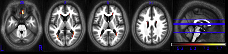
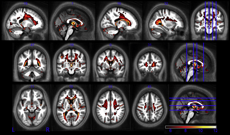
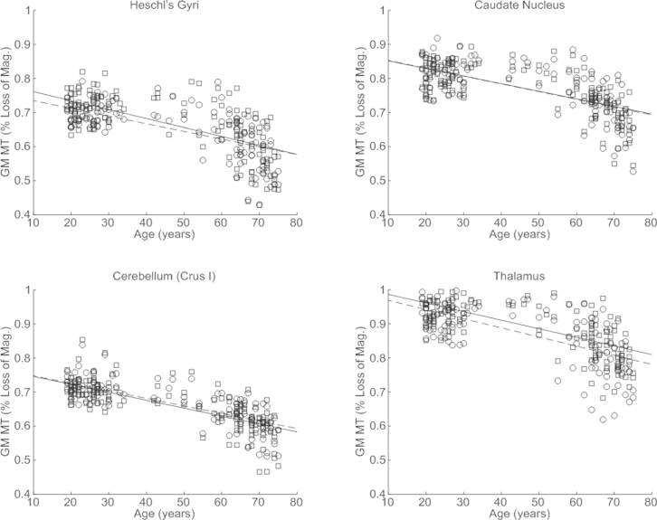
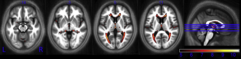
A pressing need exists to disentangle age-related changes from pathologic neurodegeneration. This study aims to characterize the spatial pattern and age-related differences of biologically relevant measures in vivo over the course of normal aging. Quantitative multiparameter maps that provide neuroimaging biomarkers for myelination and iron levels, parameters sensitive to aging, were acquired from 138 healthy volunteers (age range: 19-75 years). Whole-brain voxel-wise analysis revealed a global pattern of age-related degeneration. Significant demyelination occurred principally in the white matter. The observed age-related differences in myelination were anatomically specific. In line with invasive histologic reports, higher age-related differences were seen in the genu of the corpus callosum than the splenium. Iron levels were significantly increased in the basal ganglia, red nucleus, and extensive cortical regions but decreased along the superior occipitofrontal fascicle and optic radiation. This whole-brain pattern of age-associated microstructural differences in the asymptomatic population provides insight into the neurobiology of aging. The results help build a quantitative baseline from which to examine and draw a dividing line between healthy aging and pathologic neurodegeneration.
迫切需要将与年龄相关的变化与病理性神经退行性变区分开来。本研究旨在描述正常衰老过程中体内生物学相关指标的空间模式和与年龄相关的差异。从138名健康志愿者(年龄范围:19 - 75岁)获取了提供髓鞘形成和铁水平神经影像学生物标志物的定量多参数图,这些参数对衰老敏感。全脑体素级分析揭示了与年龄相关的退化的整体模式。显著的脱髓鞘主要发生在白质。观察到的与年龄相关的髓鞘形成差异具有解剖学特异性。与侵入性组织学报告一致,胼胝体膝部比压部出现更高的与年龄相关的差异。基底神经节、红核和广泛的皮质区域铁水平显著升高,但沿枕额上束和视辐射铁水平降低。无症状人群中这种与年龄相关的微观结构差异的全脑模式为衰老的神经生物学提供了见解。这些结果有助于建立一个定量基线,据此来检查并划分健康衰老和病理性神经退行性变之间的界限。