Issan Yossi, Kornowski Ran, Aravot Dan, Shainberg Asher, Laniado-Schwartzman Michal, Sodhi Komal, Abraham Nader G, Hochhauser Edith
Cardiac Research Laboratory, Felsenstein Medical Research Institute, Tel-Aviv University, Petah-Tikva, Israel.
Cardiac Research Laboratory, Felsenstein Medical Research Institute, Tel-Aviv University, Petah-Tikva, Israel; Cardiology Department, Rabin Medical Center, Tel-Aviv University, Petah-Tikva, Israel.
PLoS One. 2014 Mar 21;9(3):e92246. doi: 10.1371/journal.pone.0092246. eCollection 2014.
Oxidative stress plays a key role in exacerbating diabetes and cardiovascular disease. Heme oxygenase-1 (HO-1), a stress response protein, is cytoprotective, but its role in post myocardial infarction (MI) and diabetes is not fully characterized. We aimed to investigate the protection and the mechanisms of HO-1 induction in cardiomyocytes subjected to hypoxia and in diabetic mice subjected to LAD ligation.
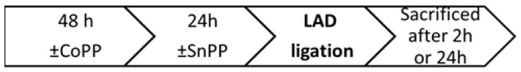
In vitro: cultured cardiomyocytes were treated with cobalt-protoporphyrin (CoPP) and tin protoporphyrin (SnPP) prior to hypoxic stress. In vivo: CoPP treated streptozotocin-induced diabetic mice were subjected to LAD ligation for 2/24 h. Cardiac function, histology, biochemical damage markers and signaling pathways were measured.
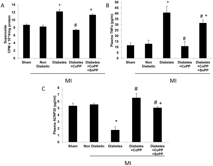
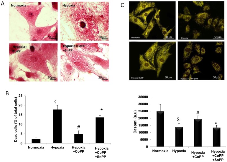
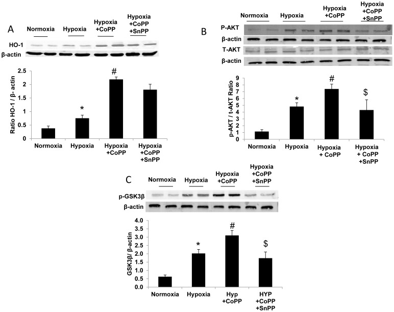
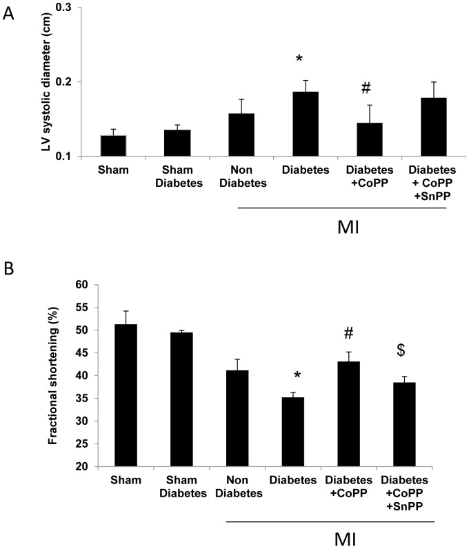
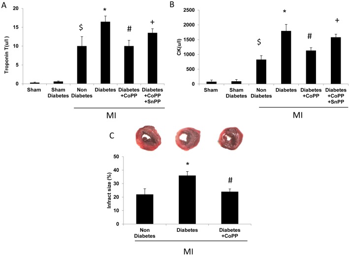
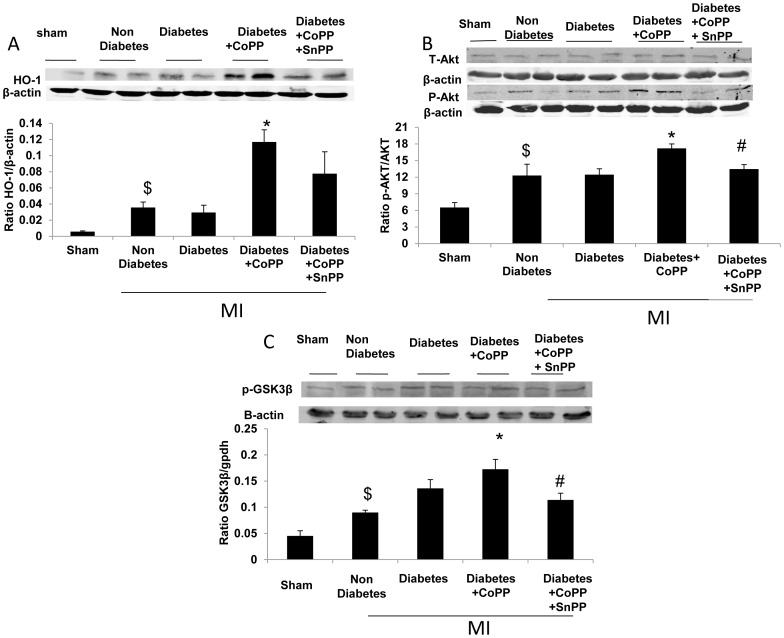
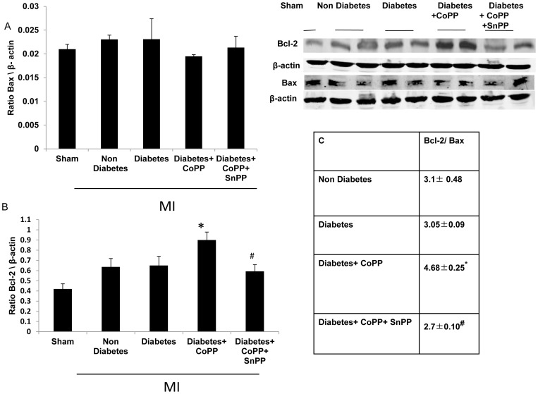
HO-1 induction lowered release of lactate dehydrogenase (LDH) and creatine phospho kinase (CK), decreased propidium iodide staining, improved cell morphology and preserved mitochondrial membrane potential in cardiomyocytes. In diabetic mice, Fractional Shortening (FS) was lower than non-diabetic mice (35±1%vs.41±2, respectively p<0.05). CoPP-treated diabetic animals improved cardiac function (43±2% p<0.01), reduced CK, Troponin T levels and infarct size compared to non-treated diabetic mice (P<0.01, P<0.001, P<0.01 respectively). CoPP-enhanced HO-1 protein levels and reduced oxidative stress in diabetic animals, as indicated by the decrease in superoxide levels in cardiac tissues and plasma TNFα levels (p<0.05). The increased levels of HO-1 by CoPP treatment after LAD ligation led to a shift of the Bcl-2/bax ratio towards the antiapoptotic process (p<0.05). CoPP significantly increased the expression levels of pAKT and pGSK3β (p<0.05) in cardiomyocytes and in diabetic mice with MI. SnPP abolished CoPP's cardioprotective effects.
HO-1 induction plays a role in cardioprotection against hypoxic damage in cardiomyocytes and in reducing post ischemic cardiac damage in the diabetic heart as proved by the increased levels of pAKT with a concomitant inhibition of pGSK3β leading to preserved mitochondrial membrane potential.
氧化应激在加重糖尿病和心血管疾病方面起关键作用。血红素加氧酶-1(HO-1)是一种应激反应蛋白,具有细胞保护作用,但其在心肌梗死(MI)后及糖尿病中的作用尚未完全明确。我们旨在研究HO-1诱导在缺氧心肌细胞及糖尿病小鼠左冠状动脉结扎模型中的保护作用及其机制。
体外实验:在缺氧应激前,用钴原卟啉(CoPP)和锡原卟啉(SnPP)处理培养的心肌细胞。体内实验:用CoPP处理链脲佐菌素诱导的糖尿病小鼠,然后进行左冠状动脉结扎2/24小时。检测心脏功能、组织学、生化损伤标志物及信号通路。
诱导HO-1可降低心肌细胞中乳酸脱氢酶(LDH)和肌酸磷酸激酶(CK)的释放,减少碘化丙啶染色,改善细胞形态并维持线粒体膜电位。在糖尿病小鼠中,短轴缩短率(FS)低于非糖尿病小鼠(分别为35±1%对41±2,p<0.05)。与未处理的糖尿病小鼠相比,CoPP处理的糖尿病动物心脏功能改善(43±2%,p<0.01),CK、肌钙蛋白T水平及梗死面积降低(分别为P<0.01、P<0.001、P<0.01)。CoPP提高了糖尿病动物中HO-1蛋白水平并降低了氧化应激,这表现为心脏组织中超氧化物水平及血浆肿瘤坏死因子α水平降低(p<0.05)。左冠状动脉结扎后经CoPP处理使HO-1水平升高,导致Bcl-2/bax比值向抗凋亡方向转变(p<0.05)。CoPP显著提高了心肌细胞及心肌梗死糖尿病小鼠中pAKT和pGSK3β的表达水平(p<0.05)。SnPP消除了CoPP的心脏保护作用。
诱导HO-1在保护心肌细胞免受缺氧损伤及减少糖尿病心脏缺血后心脏损伤中发挥作用,pAKT水平升高同时抑制pGSK3β导致线粒体膜电位维持可证明这一点。