Yoritsune Erina, Furuse Motomasa, Kuwabara Hiroko, Miyata Tomo, Nonoguchi Naosuke, Kawabata Shinji, Hayasaki Hana, Kuroiwa Toshihiko, Ono Koji, Shibayama Yuro, Miyatake Shin-Ichi
Department of Neurosurgery, Division of Life Sciences, Osaka Medical College, 2-7 Daigaku-machi, Takatsuki City, Osaka 569-8686, Japan.
Department of Pathology, Division of Life Sciences, Osaka Medical College, 2-7 Daigaku-machi, Takatsuki City, Osaka 569-8686, Japan.
J Radiat Res. 2014 Jul;55(4):803-11. doi: 10.1093/jrr/rru017. Epub 2014 Mar 27.
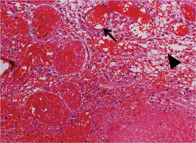
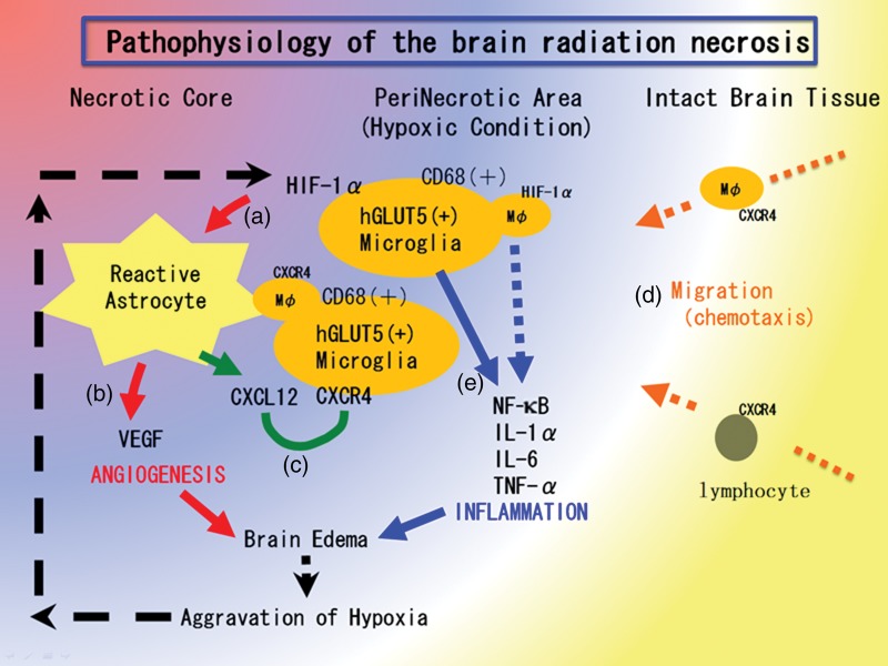
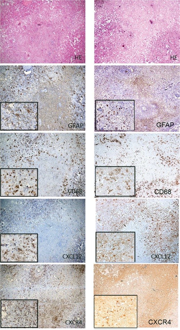
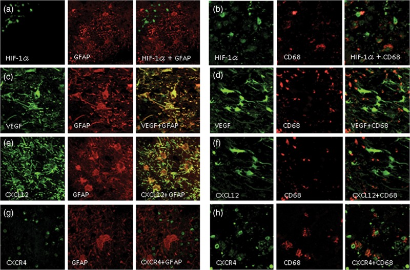
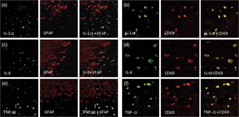
Radiation necrosis (RN) after intensive radiation therapy is a serious problem. Using human RN specimens, we recently proved that leaky angiogenesis is a major cause of brain edema in RN. In the present study, we investigated the same specimens to speculate on inflammation's effect on the pathophysiology of RN. Surgical specimens of symptomatic RN in the brain were retrospectively reviewed by histological and immunohistochemical analyses using hematoxylin and eosin (H&E) staining as well as immunohistochemical staining for VEGF, HIF-1α, CXCL12, CXCR4, GFAP, CD68, hGLUT5, CD45, IL-1α, IL-6 TNF-α and NF-kB. H&E staining demonstrated marked angiogenesis and cell infiltration in the perinecrotic area. The most prominent vasculature was identified as thin-walled leaky angiogenesis, i.e. telangiectasis surrounded by prominent interstitial edema. Two major cell phenotypes infiltrated the perinecrotic area: GFAP-positive reactive astrocytes and CD68/hGLUT5-positive cells (mainly microglias). Immunohistochemistry revealed that CD68/hGLUT5-positive cells and GFAP-positive cells expressed HIF-1α and VEGF, respectively. GFAP-positive cells expressed chemokine CXCL12, and CD68/hGLUT5-positive cells expressed receptor CXCR4. The CD68/hGLUT5-positive cells expressed pro-inflammatory cytokines IL-1α, IL-6 and TNF-α in the perinecrotic area. VEGF caused leaky angiogenesis followed by perilesional edema in RN. GFAP-positive cells expressing CXCL12 might attract CXCR4-expressing CD68/hGLUT5-positive cells into the perinecrotic area. These accumulated CD68/hGLUT5-positive cells expressing pro-inflammatory cytokines seemed to aggravate the RN edema. Both angiogenesis and inflammation might be caused by the regulation of HIF-1α, which is well known as a transactivator of VEGF and of the CXCL12/CXCR4 chemokine axis.
强化放射治疗后的放射性坏死(RN)是一个严重问题。利用人类RN标本,我们最近证实血管生成异常是RN中脑水肿的主要原因。在本研究中,我们对相同标本进行研究,以推测炎症对RN病理生理学的影响。通过苏木精和伊红(H&E)染色以及VEGF、HIF-1α、CXCL12、CXCR4、GFAP、CD68、hGLUT5、CD45、IL-1α、IL-6、TNF-α和NF-κB的免疫组化染色,对脑内有症状RN的手术标本进行回顾性组织学和免疫组化分析。H&E染色显示坏死周围区域有明显的血管生成和细胞浸润。最显著的脉管系统被确定为薄壁的血管生成异常,即被明显的间质水肿包围的毛细血管扩张。两种主要细胞表型浸润坏死周围区域:GFAP阳性的反应性星形胶质细胞和CD68/hGLUT5阳性细胞(主要是小胶质细胞)。免疫组化显示,CD68/hGLUT5阳性细胞和GFAP阳性细胞分别表达HIF-1α和VEGF。GFAP阳性细胞表达趋化因子CXCL12,CD68/hGLUT5阳性细胞表达受体CXCR4。CD68/hGLUT5阳性细胞在坏死周围区域表达促炎细胞因子IL-1α、IL-6和TNF-α。VEGF导致血管生成异常,随后在RN中出现病灶周围水肿。表达CXCL12的GFAP阳性细胞可能将表达CXCR4的CD68/hGLUT5阳性细胞吸引到坏死周围区域。这些积累的表达促炎细胞因子的CD68/hGLUT5阳性细胞似乎会加重RN水肿。血管生成和炎症可能均由HIF-1α的调节引起,HIF-1α是众所周知的VEGF以及CXCL12/CXCR4趋化因子轴的反式激活因子。