Satoh Daisuke, Hirose Tomonori, Harita Yutaka, Daimon Chikara, Harada Tomonori, Kurihara Hidetake, Yamashita Akio, Ohno Shigeo
Department of Molecular Biology, Graduate School of Medical Science, Yokohama City University, Yokohama; Department of Pediatrics, Graduate School of Medicine, The University of Tokyo, Tokyo; Department of Pediatrics, Yokohama City University, Yokohama; Department of Anatomy, Juntendo University, School of Medicine, Bunkyo, Tokyo; and Advanced Medical Research Center, Yokohama City University, Yokohama, Kanagawa, Japan.
Department of Molecular Biology, Graduate School of Medical Science, Yokohama City University, Yokohama; Department of Pediatrics, Graduate School of Medicine, The University of Tokyo, Tokyo; Department of Pediatrics, Yokohama City University, Yokohama; Department of Anatomy, Juntendo University, School of Medicine, Bunkyo, Tokyo; and Advanced Medical Research Center, Yokohama City University, Yokohama, Kanagawa, JapanDepartment of Molecular Biology, Graduate School of Medical Science, Yokohama City University, Yokohama; Department of Pediatrics, Graduate School of Medicine, The University of Tokyo, Tokyo; Department of Pediatrics, Yokohama City University, Yokohama; Department of Anatomy, Juntendo University, School of Medicine, Bunkyo, Tokyo; and Advanced Medical Research Center, Yokohama City University, Yokohama, Kanagawa, Japan.
J Biochem. 2014 Aug;156(2):115-28. doi: 10.1093/jb/mvu022. Epub 2014 Apr 3.
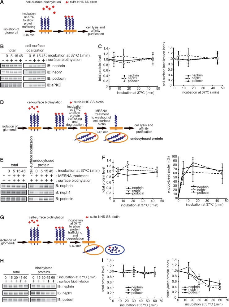
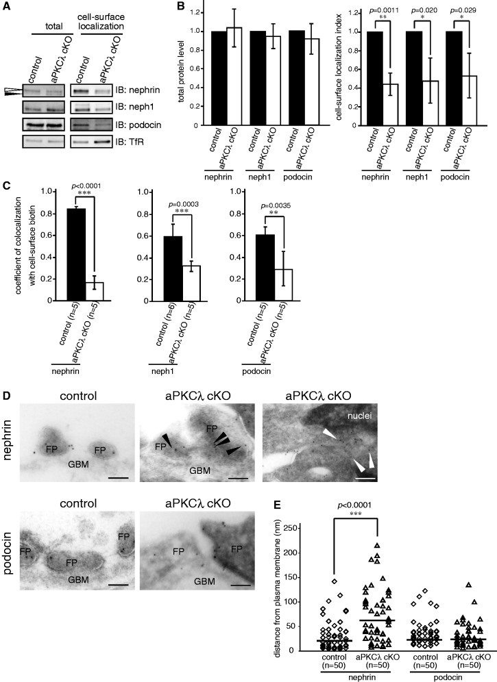
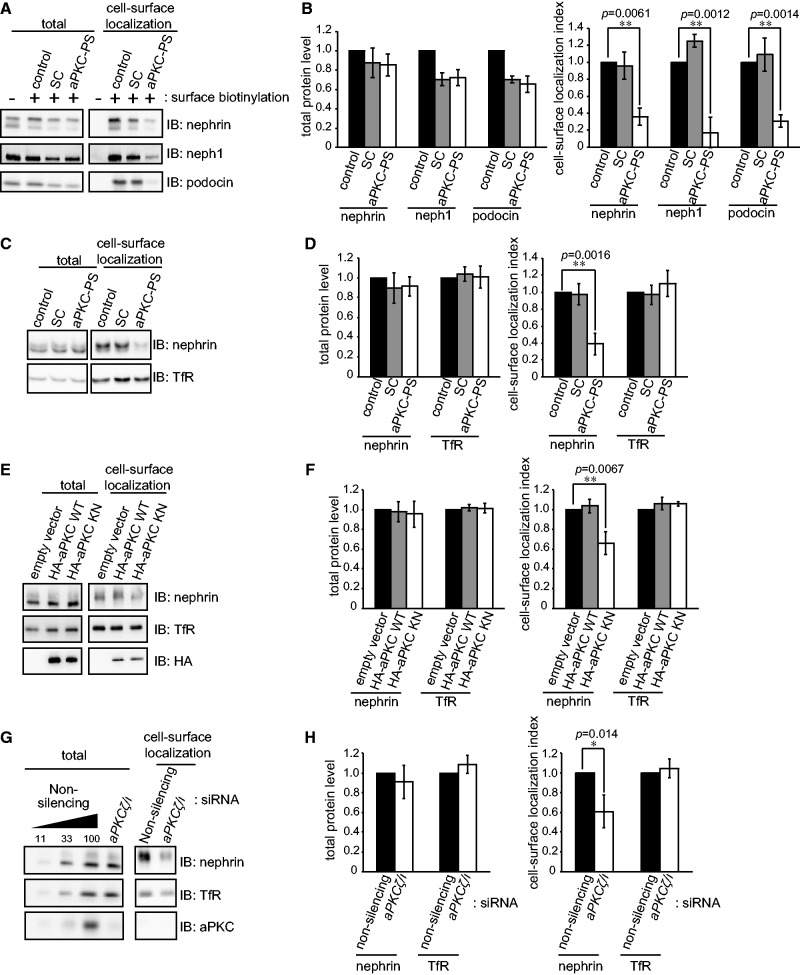
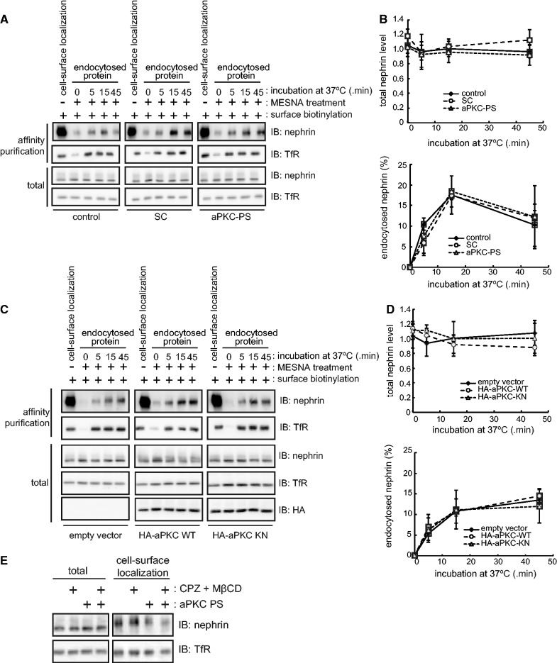
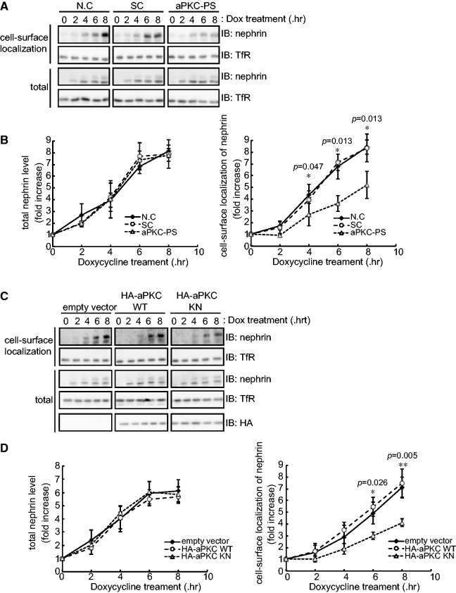
The slit diaphragm (SD), the specialized intercellular junction between renal glomerular epithelial cells (podocytes), provides a selective-filtration barrier in renal glomeruli. Dysfunction of the SD results in glomerular diseases that are characterized by disappearance of SD components, such as nephrin, from the cell surface. Although the importance of endocytosis and degradation of SD components for the maintenance of SD integrity has been suggested, the dynamic nature of the turnover of intact cell-surface SD components remained unclear. Using isolated rat glomeruli we show that the turnover rates of cell-surface SD components are relatively high; they almost completely disappear from the cell surface within minutes. The exocytosis, but not endocytosis, of heterologously expressed nephrin requires the kinase activity of the cell polarity regulator atypical protein kinase C (aPKC). Consistently, we demonstrate that podocyte-specific deletion of aPKCλ resulted in a decrease of cell-surface localization of SD components, causing massive proteinuria. In conclusion, the regulation of SD turnover by aPKC is crucial for the maintenance of SD integrity and defects in aPKC signalling can lead to proteinuria. These findings not only reveal the pivotal importance of the dynamic turnover of cell-surface SD components but also suggest a novel pathophysiological basis in glomerular disease.
裂孔隔膜(SD)是肾肾小球上皮细胞(足细胞)之间的特殊细胞间连接,在肾肾小球中提供选择性滤过屏障。SD功能障碍会导致肾小球疾病,其特征是SD成分(如nephrin)从细胞表面消失。尽管内吞作用和SD成分降解对维持SD完整性的重要性已被提出,但完整细胞表面SD成分周转的动态性质仍不清楚。我们使用分离的大鼠肾小球表明,细胞表面SD成分的周转率相对较高;它们在几分钟内几乎完全从细胞表面消失。异源表达的nephrin的胞吐作用而非内吞作用需要细胞极性调节剂非典型蛋白激酶C(aPKC)的激酶活性。一致地,我们证明足细胞特异性缺失aPKCλ会导致SD成分的细胞表面定位减少,从而导致大量蛋白尿。总之,aPKC对SD周转的调节对于维持SD完整性至关重要,aPKC信号缺陷可导致蛋白尿。这些发现不仅揭示了细胞表面SD成分动态周转的关键重要性,还提示了肾小球疾病的新病理生理基础。