Department of Internal Medicine, Gastroenterology and Hepatology, University Hospital, University of Warmia and Mazury, Olsztyn, Poland ; Department of Internal Medicine, Cardiology and Nephrology, Hospital, Ostroda, Poland.
Department of Internal Medicine and Cardiology, Municipal Hospital, Olsztyn, Poland.
Arch Med Sci. 2014 Feb 24;10(1):39-46. doi: 10.5114/aoms.2014.40732. Epub 2014 Feb 23.
The diagnosis of acute pulmonary embolism (APE) in patients with chronic heart failure (CHF) remains a difficult task, despite the refinement of imaging techniques. The goal of this study was to assess the value of measuring tricuspid and mitral valve systolic annular velocities in CHF patients with suspected PE by tissue Doppler imaging (TDI).
The study included 75 patients with previously diagnosed CHF, admitted due to resting dyspnea, with a maximum tricuspid regurgitation pressure gradient (TRPG) of ≥ 35 mm Hg and positive D-dimer assay. Spiral computed tomography (sCT) was performed on all subjects to confirm APE. Acute pulmonary embolism was diagnosed in 35 patients (PE+), and excluded in 40 others (PE-). Tissue Doppler imaging was performed to measure maximum systolic lateral annular velocities in the mitral (SmLV) and tricuspid (SmRV) valves, as well as the SmRV/SmLV ratio.
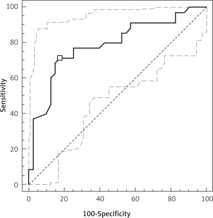
PE+ subjects were found to have higher SmLV than PE- subjects (6.0 cm/s (2.0-13.8 cm/s) vs. 4.2 cm/s (1.3-9.1 cm/s), p = 0.003). SmRV/SmLV ratios were 1.05 (0.50-2.50) and 1.56 (0.62-4.30), respectively (p < 0.0001). Areas under ROC curves for diagnosis of APE were 0.700 for SmLV and 0.789 for SmRV/SmLV. In multivariate logistic regression analysis, only SmRV/SmLV was statistically significant, with an odds ratio for APE of 6.26 (95% CI: 1.53-25.59; p = 0.009).
Tissue Doppler imaging of the lateral tricuspid and mitral annuli is a useful clinical tool that can help identify PE in CHF patients. Those patients who fulfill these criteria should be considered for further diagnostic studies to confirm PE.
尽管影像学技术不断发展,但在慢性心力衰竭(CHF)患者中诊断急性肺栓塞(APE)仍然是一项艰巨的任务。本研究旨在通过组织多普勒成像(TDI)评估测量疑似 PE 的 CHF 患者三尖瓣和二尖瓣收缩环速度的价值。
该研究纳入了 75 名先前诊断为 CHF 的患者,因静息性呼吸困难入院,最大三尖瓣反流压差(TRPG)≥35mmHg,D-二聚体检测阳性。所有患者均行螺旋 CT(sCT)检查以确诊 APE。35 例患者(PE+)确诊为 APE,40 例患者(PE-)排除 APE。通过组织多普勒成像测量二尖瓣(SmLV)和三尖瓣(SmRV)收缩环的最大侧环速度以及 SmRV/SmLV 比值。
PE+患者的 SmLV 高于 PE-患者(6.0cm/s[2.0-13.8cm/s] vs. 4.2cm/s[1.3-9.1cm/s],p=0.003)。SmRV/SmLV 比值分别为 1.05(0.50-2.50)和 1.56(0.62-4.30)(p<0.0001)。SmLV 和 SmRV/SmLV 诊断 APE 的 ROC 曲线下面积分别为 0.700 和 0.789。多变量逻辑回归分析显示,仅 SmRV/SmLV 具有统计学意义,APE 的优势比为 6.26(95%CI:1.53-25.59;p=0.009)。
三尖瓣和二尖瓣侧环的组织多普勒成像技术是一种有用的临床工具,可帮助识别 CHF 患者中的 PE。符合这些标准的患者应考虑进一步进行诊断研究以确诊 PE。