Cabibi D, Giannone A G, Mascarella C, Guarnotta C, Castiglia M, Pantuso G, Fiorentino E
Department of Sciences for Promotion of Health and Mother and Child care - University of Palermo.
Eur J Histochem. 2014 Mar 5;58(1):2326. doi: 10.4081/ejh.2014.2326.
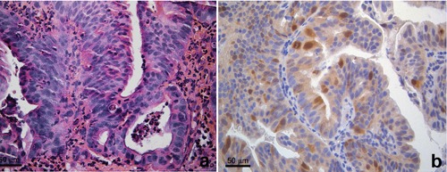
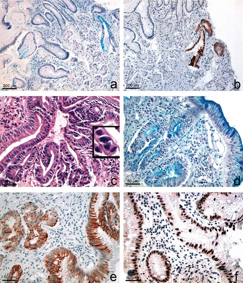
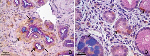
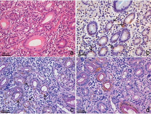
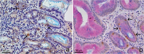
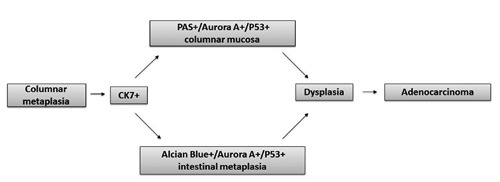
Intestinal metaplasia in Barrett's oesophagus (BO) represents an important risk factor for oesophageal adenocarcinoma. Instead, few and controversial data are reported about the progression risk of columnar-lined oesophagus without intestinal metaplasia (CLO), posing an issue about its clinical management. The aim was to evaluate if some immunophenotypic changes were present in CLO independently of the presence of the goblet cells. We studied a series of oesophageal biopsies from patients with endoscopic finding of columnar metaplasia, by performing some immunohistochemical stainings (CK7, p53, AuroraA) combined with histochemistry (Alcian-blue and Alcian/PAS), with the aim of simultaneously assess the histochemical features in cells that shows an aberrant expression of such antigens. We evidenced a cytoplasmic expression of CK7 and a nuclear expression of Aurora A and p53, both in goblet cells of BO and in non-goblet cells of CLO, some of which showing mild dysplasia. These findings suggest that some immunophenotypic changes are present in CLO and they can precede the appearance of the goblet cells or can be present independently of them, confirming the conception of BO as the condition characterized by any extention of columnar epithelium. This is the first study in which a combined immunohistochemical/histochemical method has been applied to Barrett pathology.
巴雷特食管(BO)中的肠化生是食管腺癌的一个重要危险因素。相反,关于无肠化生的柱状上皮食管(CLO)进展风险的报道很少且存在争议,这对其临床管理提出了一个问题。目的是评估CLO中是否存在一些免疫表型变化,而不考虑杯状细胞的存在。我们通过进行一些免疫组织化学染色(CK7、p53、AuroraA)并结合组织化学(阿尔辛蓝和阿尔辛蓝/过碘酸雪夫染色),研究了一系列经内镜检查发现柱状化生患者的食管活检组织,目的是同时评估显示此类抗原异常表达的细胞中的组织化学特征。我们发现,在BO的杯状细胞和CLO的非杯状细胞中均有CK7的细胞质表达以及Aurora A和p53的核表达,其中一些显示轻度发育异常。这些发现表明,CLO中存在一些免疫表型变化,它们可能先于杯状细胞出现,或者可以独立于杯状细胞存在,这证实了将BO视为以柱状上皮任何延伸为特征的疾病的概念。这是第一项将免疫组织化学/组织化学联合方法应用于巴雷特病理学的研究。