Zhang Yijie, Zheng Kailian, Yan Hongli, Jin Gang, Shao Chenghao, Zhou Xuyu, Zhou Yingqi, He Tianlin
Department of General Surgery, Changhai Hospital, No,168 Changhai Road, Shanghai, Yangpu District 200433, China.
BMC Gastroenterol. 2014 Apr 3;14:62. doi: 10.1186/1471-230X-14-62.
Hepatocellular carcinoma (HCC) is one of the most lethal and prevalent cancers in human population. The 6-fluoro-3-formylchromone (FCC) has been shown to have anti-tumor activity against various tumor cells. However, the effects of FCC on HCC cell lines have not yet been reported. This study aims to research the effects of FCC on HCC and advance the understanding of the molecular mechanism.
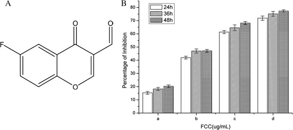
HCC cell line SMMC-7721 was treated with FCC at various concentrations (0, 2, 5, 10, and 20 μg/ml) for 24, 48 and 72 h, respectively. The proliferations of SMMC-7721 cells were measured by MTT assays. After cultured 24 hours, cell cycle distribution and apoptosis were determined by flow cytometry. However, the expression levels of PCNA, Bax and Bcl-2 were measured by western blotting after 48 hours.
FCC displayed a dose- and time-dependent inhibition of the SMMC-7721 cell proliferations in vitro. It also induced apoptosis with 45.4% and caused cell accumulation in G0/G1 phase with 21.5%. PCNA and Bcl-2 expression was significantly suppressed by FCC in a dose-dependent manner (P < 0.05), while Bax expression was increased.
FCC could significantly inhibit HCC cell growth in vitro through cell cycle arrest and inducing apoptosis by suppressing PCNA expression and modulating the Bax/Bcl-2 ratio.
肝细胞癌(HCC)是人类最致命且常见的癌症之一。6-氟-3-甲酰基色酮(FCC)已显示出对多种肿瘤细胞具有抗肿瘤活性。然而,FCC对肝癌细胞系的影响尚未见报道。本研究旨在探究FCC对肝癌的影响,并加深对其分子机制的理解。
分别用不同浓度(0、2、5、10和20μg/ml)的FCC处理肝癌细胞系SMMC-7721,处理时间分别为24、48和72小时。通过MTT法检测SMMC-7721细胞的增殖情况。培养24小时后,通过流式细胞术测定细胞周期分布和凋亡情况。然而,48小时后通过蛋白质印迹法检测PCNA、Bax和Bcl-2的表达水平。
FCC在体外对SMMC-7721细胞的增殖具有剂量和时间依赖性抑制作用。它还诱导了45.4%的细胞凋亡,并使21.5%的细胞停滞在G0/G1期。FCC以剂量依赖性方式显著抑制PCNA和Bcl-2的表达(P<0.05),而Bax的表达增加。
FCC可通过抑制PCNA表达和调节Bax/Bcl-2比值,使细胞周期停滞并诱导凋亡,从而在体外显著抑制肝癌细胞生长。