Frank Reidy Research Center for Bioelectrics, Old Dominion University, 4211 Monarch Way, IRP2, Norfolk, Virginia, 23508, USA.
Division of Molecular Carcinogenesis and Targeted Therapy for Cancer, Chinese Academy of Sciences, 1 Beichen West Road, Beijing 100101, China.
Cells. 2013 Mar 6;2(1):136-62. doi: 10.3390/cells2010136.
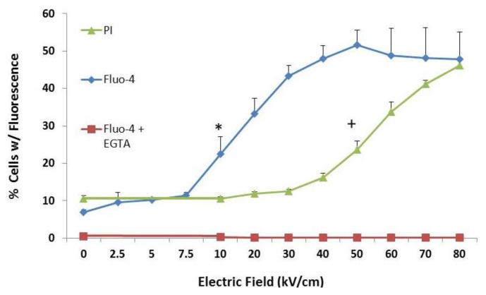
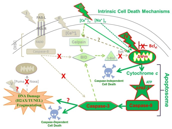
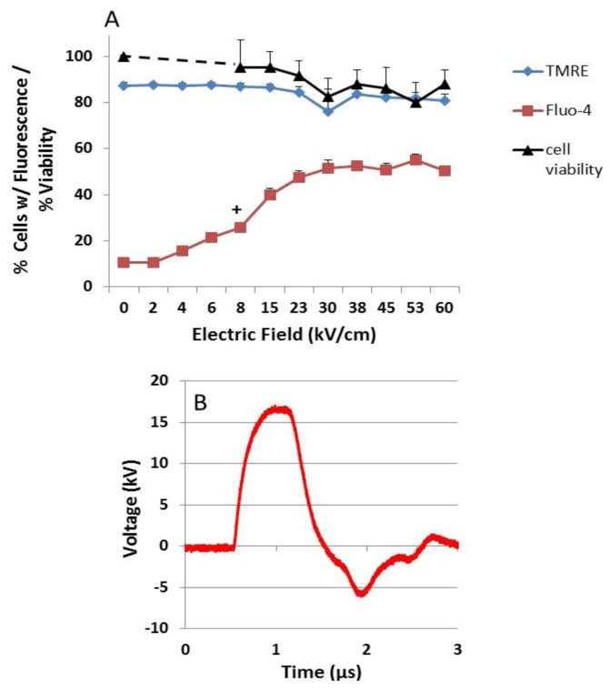
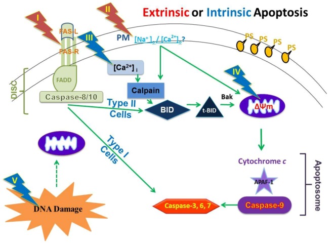
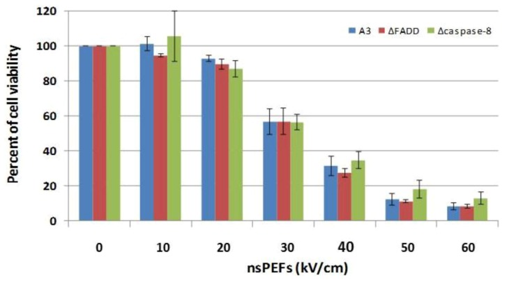
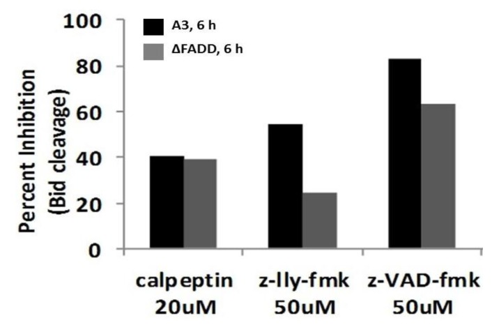
Pulse power technology using nanosecond pulsed electric fields (nsPEFs) offers a new stimulus to modulate cell functions or induce cell death for cancer cell ablation. New data and a literature review demonstrate fundamental and basic cellular mechanisms when nsPEFs interact with cellular targets. NsPEFs supra-electroporate cells creating large numbers of nanopores in all cell membranes. While nsPEFs have multiple cellular targets, these studies show that nsPEF-induced dissipation of ΔΨm closely parallels deterioration in cell viability. Increases in intracellular Ca2+ alone were not sufficient for cell death; however, cell death depended of the presence of Ca2+. When both events occur, cell death ensues. Further, direct evidence supports the hypothesis that pulse rise-fall times or high frequency components of nsPEFs are important for decreasing ΔΨm and cell viability. Evidence indicates in Jurkat cells that cytochrome c release from mitochondria is caspase-independent indicating an absence of extrinsic apoptosis and that cell death can be caspase-dependent and -independent. The Ca2+ dependence of nsPEF-induced dissipation of ΔΨm suggests that nanoporation of inner mitochondria membranes is less likely and effects on a Ca2+-dependent protein(s) or the membrane in which it is embedded are more likely a target for nsPEF-induced cell death. The mitochondria permeability transition pore (mPTP) complex is a likely candidate. Data demonstrate that nsPEFs can bypass cancer mutations that evade apoptosis through mechanisms at either the DISC or the apoptosome.
利用纳秒脉冲电场(nsPEFs)的脉冲功率技术为调节细胞功能或诱导癌细胞消融提供了一种新的刺激。新的数据和文献综述表明,当 nsPEFs 与细胞靶标相互作用时,存在基本的细胞机制。nsPEFs 超电穿孔细胞,在所有细胞膜上产生大量纳米孔。虽然 nsPEFs 有多个细胞靶标,但这些研究表明,nsPEF 诱导的ΔΨm 耗散与细胞活力的恶化密切相关。单独增加细胞内 Ca2+ 不足以引起细胞死亡;然而,细胞死亡取决于 Ca2+ 的存在。当这两种情况同时发生时,细胞死亡就会发生。此外,直接证据支持这样的假设,即 nsPEF 的脉冲上升-下降时间或高频成分对于降低ΔΨm 和细胞活力很重要。证据表明,在 Jurkat 细胞中,细胞色素 c 从线粒体中的释放是 caspase 非依赖性的,表明不存在外在凋亡,并且细胞死亡可以是 caspase 依赖性和非依赖性的。nsPEF 诱导的ΔΨm 耗散的 Ca2+依赖性表明,内线粒体膜的纳米孔形成不太可能,而对 Ca2+-依赖性蛋白(或其嵌入的膜)的影响更可能是 nsPEF 诱导细胞死亡的靶标。线粒体通透性转换孔(mPTP)复合物是一个可能的候选物。数据表明,nsPEFs 可以绕过通过 DISC 或凋亡小体中的机制逃避细胞凋亡的癌症突变。