Nagoya University Graduate School of Medicine, Nagoya Kyoritsu Hospital.
J Appl Clin Med Phys. 2014 Mar 6;15(2):4603. doi: 10.1120/jacmp.v15i2.4603.
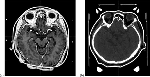
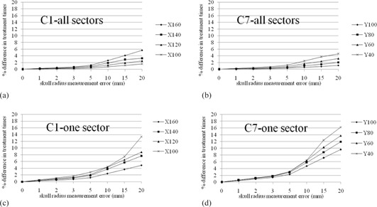
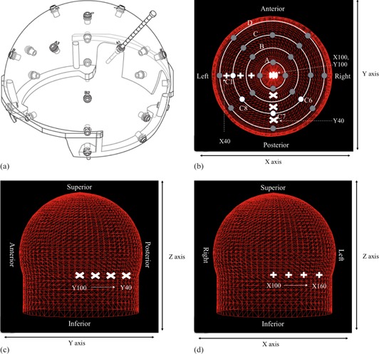
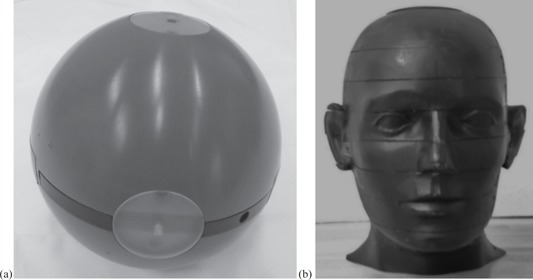
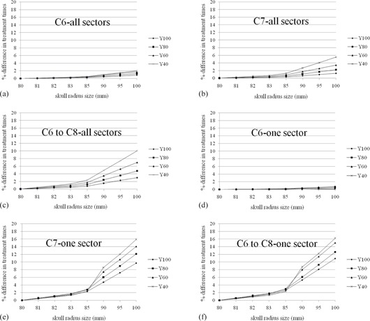
In treatment planning of Leksell Gamma Knife (LGK) radiosurgery, the skull geometry defined by generally dedicated scalar measurement has a crucial effect on dose calculation. The LGK Perfexion (PFX) unit is equipped with a cone-shaped collimator divided into eight sectors, and its configuration is entirely different from previous model C. Beam delivery on the PFX is made by a combination of eight sectors, but it is also mechanically available from one sector with the remaining seven blocked. Hence the treatment time using one sector is more likely to be affected by discrepancies in the skull shape than that of all sectors. In addition, the latest version (Ver. 10.1.1) of the treatment planning system Leksell GammaPlan (LGP) includes a new function to directly generate head surface contouring from computed tomography (CT) images in conjunction with the Leksell skull frame. This paper evaluates change of treatment time induced by different skull models. A simple simulation using a uniform skull radius of 80 mm and anthropomorphic phantom was implemented in LGP to find the trend between dose and skull measuring error. To evaluate the clinical effect, we performed an interobserver comparison of ruler measurement for 41 patients, and compared instrumental and CT-based contours for 23 patients. In the phantom simulation, treatment time errors were less than 2% when the difference was within 3 mm. In the clinical cases, the variability of treatment time induced by the differences in interobserver measurements was less than 0.91%, on average. Additionally the difference between measured and CT-based contours was good, with a difference of -0.16% ± 0.66% (mean ±1 standard deviation) on average and a maximum of 3.4%. Although the skull model created from CT images reduced the dosimetric uncertainty caused by different measurers, these results showed that even manual skull measurement could reproduce the skull shape close to that of a patient's head within an acceptable range.
在 Leksell Gamma Knife(LGK)伽玛刀放射外科治疗计划中,通常专用标量测量定义的颅骨几何形状对剂量计算有至关重要的影响。LGK Perfexion(PFX)单元配备了一个锥形准直器,分为八个扇区,其配置与以前的 C 型完全不同。PFX 的束流传输由八个扇区组合而成,但也可以通过机械方式阻挡其余七个扇区中的一个扇区来实现。因此,使用一个扇区进行治疗的时间比使用所有扇区更容易受到颅骨形状差异的影响。此外,最新版本(版本 10.1.1)的治疗计划系统 Leksell GammaPlan(LGP)包括一个新功能,可直接从 CT 图像生成头表面轮廓,并结合 Leksell 颅骨框架。本文评估了不同颅骨模型引起的治疗时间变化。在 LGP 中使用均匀颅骨半径 80mm 和人体模型进行了简单的模拟,以找到剂量和颅骨测量误差之间的趋势。为了评估临床效果,我们对 41 名患者进行了尺子测量的观察者间比较,并对 23 名患者进行了仪器和 CT 基轮廓的比较。在体模模拟中,当差异在 3mm 以内时,治疗时间误差小于 2%。在临床病例中,观察者间测量差异引起的治疗时间变化的可变性平均小于 0.91%。此外,测量和 CT 基轮廓之间的差异也很好,平均差异为-0.16%±0.66%(平均值±1 个标准差),最大差异为 3.4%。尽管 CT 图像生成的颅骨模型减少了不同测量者引起的剂量不确定性,但这些结果表明,即使是手动颅骨测量也可以在可接受的范围内再现接近患者头部的颅骨形状。