• 文献检索
  • 文档翻译
  • 深度研究
  • 学术资讯
  • Suppr Zotero 插件Zotero 插件
  • 邀请有礼
  • 套餐&价格
  • 历史记录
应用&插件
Suppr Zotero 插件Zotero 插件浏览器插件Mac 客户端Windows 客户端微信小程序
定价
高级版会员购买积分包购买API积分包
服务
文献检索文档翻译深度研究API 文档MCP 服务
关于我们
关于 Suppr公司介绍联系我们用户协议隐私条款
关注我们

Suppr 超能文献

核心技术专利:CN118964589B侵权必究
粤ICP备2023148730 号-1Suppr @ 2026

文献检索

告别复杂PubMed语法,用中文像聊天一样搜索,搜遍4000万医学文献。AI智能推荐,让科研检索更轻松。

立即免费搜索

文件翻译

保留排版,准确专业,支持PDF/Word/PPT等文件格式,支持 12+语言互译。

免费翻译文档

深度研究

AI帮你快速写综述,25分钟生成高质量综述,智能提取关键信息,辅助科研写作。

立即免费体验

使用对比增强三维超声对胰腺实性病变进行鉴别诊断。

Differential diagnosis of solid pancreatic lesions using contrast-enhanced three-dimensional ultrasonography.

作者信息

Miwa Haruo, Numata Kazushi, Sugimori Kazuya, Kaneko Takashi, Sakamaki Kentaro, Ueda Michio, Fukuda Hiroyuki, Tanaka Katsuaki, Maeda Shin

机构信息

Gastroenterological Center, Yokohama City University Medical Center, 4-57 Urafune-cho, Minami-ku, Yokohama, Kanagawa, 232-0024, Japan.

出版信息

Abdom Imaging. 2014 Oct;39(5):988-99. doi: 10.1007/s00261-014-0135-8.

DOI:10.1007/s00261-014-0135-8
PMID:24711063
原文链接:https://pmc.ncbi.nlm.nih.gov/articles/PMC4168219/
Abstract

PURPOSE

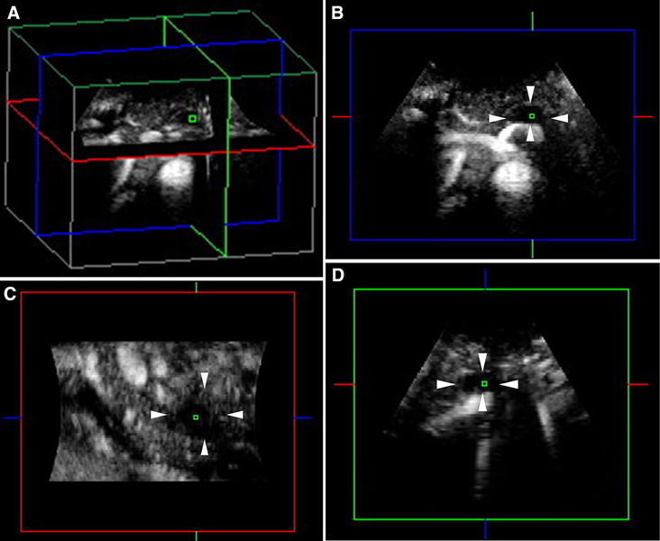
To investigate the usefulness of contrast-enhanced three-dimensional ultrasonography (CE 3D US) for differential diagnosis of solid pancreatic lesions.

METHODS

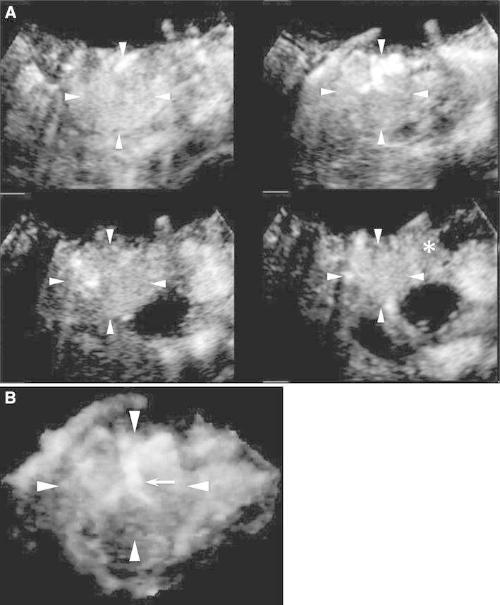
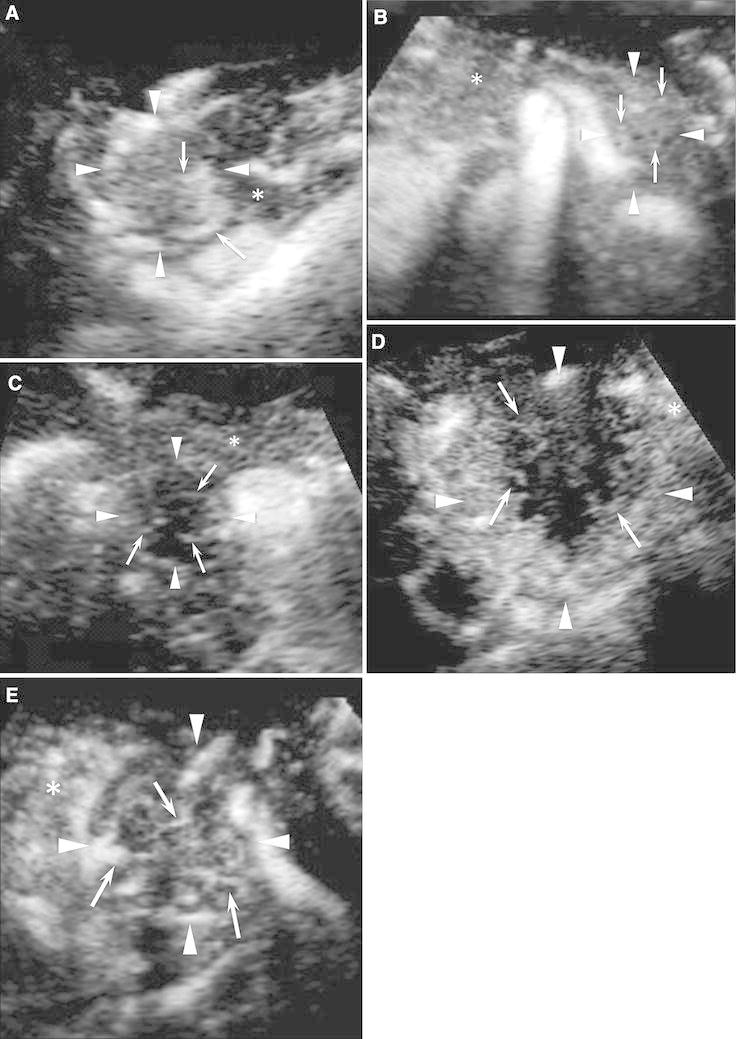
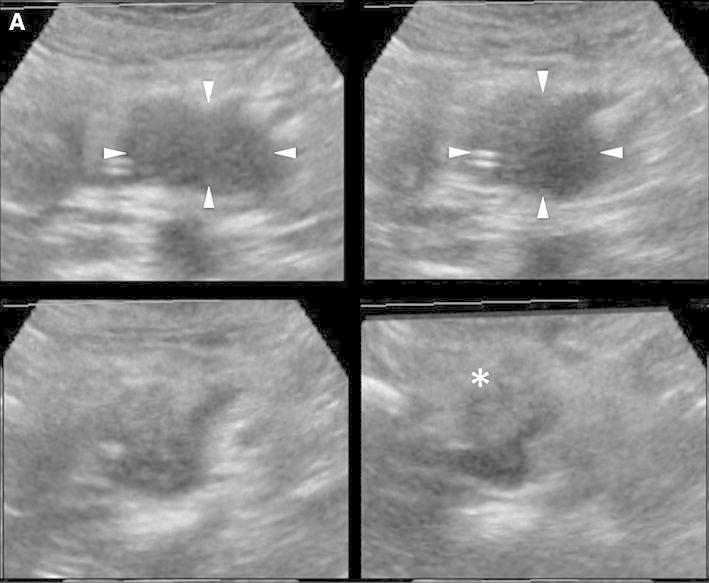
Eighty-five patients with solid pancreatic lesions who underwent CE 3D US were retrospectively analyzed. Sixty-four patients had pancreatic ductal adenocarcinoma (PDAC), 10 had mass-forming pancreatitis (MFP), and 11 had neuroendocrine tumor (NET). Two blinded readers evaluated the enhancement patterns using four features: vascularity in the arterial phase, vascularity in the venous phase, vessel location, and vessel form. Vascularity in both phases was classified as hypervascular, isovascular, or hypovascular. Vessel location was classified into peritumoral or intratumoral. Vessel form was classified into fine or irregular. Kappa values were used to assess inter-reader agreement. The institutional review board approved this study, and informed consent was obtained.

RESULTS

Kappa values of the four features were 0.75, 0.72, 0.85, and 0.65, which were graded as good or excellent. The most typical combined enhancement pattern in PDAC was hypovascularity in both phases with peritumoral and irregular vessels; MFP was isovascular in both phases with intratumoral and fine vessels; and NETs were hypervascular in both phases with intratumoral and irregular vessels. The sensitivity and positive predictive value of the three patterns were 93.8% and 96.7% for the PDAC pattern, 80.0% and 100% for the MFP pattern, and 81.8%, and 69.2% for the NET pattern, respectively. The accuracy of these diagnostic criteria was 90.5%.

CONCLUSION

CE 3D US allows detailed visualization of the enhancement patterns of various pancreatic lesions and can be used for the differential diagnosis.

摘要

目的

探讨对比增强三维超声(CE 3D US)在胰腺实性病变鉴别诊断中的应用价值。

方法

回顾性分析85例接受CE 3D US检查的胰腺实性病变患者。其中64例为胰腺导管腺癌(PDAC),10例为肿块型胰腺炎(MFP),11例为神经内分泌肿瘤(NET)。两名盲法阅片者利用四个特征评估增强模式:动脉期血管分布、静脉期血管分布、血管位置和血管形态。两期血管分布分为高血管、等血管或低血管。血管位置分为瘤周或瘤内。血管形态分为纤细或不规则。kappa值用于评估阅片者间的一致性。本研究获机构审查委员会批准,并获得了知情同意。

结果

四个特征的kappa值分别为0.75、0.72、0.85和0.65,均为良好或优秀等级。PDAC最典型的联合增强模式为两期均为低血管,伴有瘤周和不规则血管;MFP为两期均为等血管,伴有瘤内和纤细血管;NET为两期均为高血管,伴有瘤内和不规则血管。三种模式的敏感性和阳性预测值分别为:PDAC模式93.8%和96.7%,MFP模式80.0%和100%,NET模式81.8%和69.2%。这些诊断标准的准确性为90.5%。

结论

CE 3D US能够详细显示各种胰腺病变的增强模式,可用于鉴别诊断。

https://cdn.ncbi.nlm.nih.gov/pmc/blobs/33a6/4168219/4167d56afb9f/261_2014_135_Fig4_HTML.jpg
https://cdn.ncbi.nlm.nih.gov/pmc/blobs/33a6/4168219/652a235fe4ba/261_2014_135_Fig1_HTML.jpg
https://cdn.ncbi.nlm.nih.gov/pmc/blobs/33a6/4168219/cf2db78660e1/261_2014_135_Fig2a_HTML.jpg
https://cdn.ncbi.nlm.nih.gov/pmc/blobs/33a6/4168219/4a7753d0734c/261_2014_135_Fig3a_HTML.jpg
https://cdn.ncbi.nlm.nih.gov/pmc/blobs/33a6/4168219/4167d56afb9f/261_2014_135_Fig4_HTML.jpg

相似文献

1
Differential diagnosis of solid pancreatic lesions using contrast-enhanced three-dimensional ultrasonography.使用对比增强三维超声对胰腺实性病变进行鉴别诊断。
Abdom Imaging. 2014 Oct;39(5):988-99. doi: 10.1007/s00261-014-0135-8.
2
Improved differentiation of pancreatic tumors using contrast-enhanced endoscopic ultrasound.使用对比增强内镜超声改善胰腺肿瘤的鉴别诊断
Clin Gastroenterol Hepatol. 2008 May;6(5):590-597.e1. doi: 10.1016/j.cgh.2008.02.030.
3
Contrast-Enhanced Endoscopic Ultrasonography for Pancreatic Tumors.对比增强内镜超声检查在胰腺肿瘤中的应用
Biomed Res Int. 2015;2015:491782. doi: 10.1155/2015/491782. Epub 2015 May 18.
4
Differential diagnosis of pancreatic tumors using ultrasound contrast imaging.超声造影成像在胰腺肿瘤鉴别诊断中的应用
J Gastroenterol. 2005 May;40(5):518-25. doi: 10.1007/s00535-005-1578-z.
5
Clinical Value of Contrast-Enhanced Ultrasound Enhancement Patterns for Differentiating Focal Pancreatitis From Pancreatic Carcinoma: A Comparison Study With Conventional Ultrasound.超声造影增强模式在鉴别局灶性胰腺炎与胰腺癌中的临床价值:与传统超声的对比研究
J Ultrasound Med. 2018 Mar;37(3):551-559. doi: 10.1002/jum.14363. Epub 2017 Aug 29.
6
Differentiation of hypovascular pancreatic neuroendocrine tumors from pancreatic ductal adenocarcinoma using contrast-enhanced computed tomography.使用增强 CT 对乏血供胰腺神经内分泌肿瘤与胰腺导管腺癌进行鉴别。
PLoS One. 2019 Feb 1;14(2):e0211566. doi: 10.1371/journal.pone.0211566. eCollection 2019.
7
Multiphase evaluation of contrast-enhanced endoscopic ultrasonography in the diagnosis of pancreatic solid lesions.对比增强型内镜超声在胰腺实性病变诊断中的多相评估。
Pancreatology. 2018 Apr;18(3):291-297. doi: 10.1016/j.pan.2018.02.002. Epub 2018 Feb 9.
8
Characterization of small solid tumors in the pancreas: the value of contrast-enhanced harmonic endoscopic ultrasonography.胰腺小实体瘤的特征:对比增强谐波超声内镜检查的价值。
Am J Gastroenterol. 2012 Feb;107(2):303-10. doi: 10.1038/ajg.2011.354. Epub 2011 Oct 18.
9
Nonhypervascular pancreatic neuroendocrine tumors: Spectrum of MDCT imaging findings and differentiation from pancreatic ductal adenocarcinoma.非富血供胰腺神经内分泌肿瘤:MDCT 成像表现谱及与胰腺导管腺癌的鉴别诊断。
Eur J Radiol. 2019 Jan;110:66-73. doi: 10.1016/j.ejrad.2018.04.006. Epub 2018 Apr 11.
10
Nonhypervascular Pancreatic Neuroendocrine Tumors: Differential Diagnosis from Pancreatic Ductal Adenocarcinomas at MR Imaging-Retrospective Cross-sectional Study.非富血供胰腺神经内分泌肿瘤:MR 成像鉴别诊断胰腺导管腺癌——一项回顾性横断面研究。
Radiology. 2017 Jul;284(1):77-87. doi: 10.1148/radiol.2016160586. Epub 2017 Jan 13.

引用本文的文献

1
Differential Diagnosis of Solid Pancreatic Lesions Using Detective Flow Imaging Endoscopic Ultrasonography.利用检测血流成像内镜超声对胰腺实性病变进行鉴别诊断。
Diagnostics (Basel). 2024 Apr 24;14(9):882. doi: 10.3390/diagnostics14090882.
2
Anticancer Effect of Heparin-Taurocholate Conjugate on Orthotopically Induced Exocrine and Endocrine Pancreatic Cancer.肝素-牛磺胆酸盐共轭物对原位诱导的胰腺外分泌癌和内分泌癌的抗癌作用
Cancers (Basel). 2021 Nov 18;13(22):5775. doi: 10.3390/cancers13225775.
3
The AFSUMB Consensus Statements and Recommendations for the Clinical Practice of Contrast-Enhanced Ultrasound using Sonazoid.

本文引用的文献

1
Three-dimensional contrast-enhanced ultrasonography of intraductal papillary mucinous neoplasms of the pancreas: a comparison with magnetic resonance imaging.胰腺导管内乳头状黏液性肿瘤的三维对比增强超声:与磁共振成像的比较。
Pancreas. 2013 Oct;42(7):1164-8. doi: 10.1097/MPA.0b013e318291fbe5.
2
Characterization of small solid tumors in the pancreas: the value of contrast-enhanced harmonic endoscopic ultrasonography.胰腺小实体瘤的特征:对比增强谐波超声内镜检查的价值。
Am J Gastroenterol. 2012 Feb;107(2):303-10. doi: 10.1038/ajg.2011.354. Epub 2011 Oct 18.
3
Detection of pancreatic carcinoma and liver metastases with gadoxetic acid-enhanced MR imaging: comparison with contrast-enhanced multi-detector row CT.
AFSUMB关于使用声诺维进行超声造影临床实践的共识声明与建议。
J Med Ultrasound. 2020 May 25;28(2):59-82. doi: 10.4103/JMU.JMU_124_19. eCollection 2020 Apr-Jun.
4
The AFSUMB Consensus Statements and Recommendations for the Clinical Practice of Contrast-Enhanced Ultrasound using Sonazoid.AFSUMB关于使用声诺维进行超声造影临床实践的共识声明与建议。
Ultrasonography. 2020 Jul;39(3):191-220. doi: 10.14366/usg.20057. Epub 2020 Apr 27.
5
Vascular evaluation using transabdominal ultrasound for gallbladder polyps.经腹部超声对胆囊息肉的血管评估。
J Med Ultrason (2001). 2021 Apr;48(2):159-173. doi: 10.1007/s10396-020-01008-8. Epub 2020 Mar 3.
6
Differentiation of pancreatic neuroendocrine carcinoma from pancreatic ductal adenocarcinoma using magnetic resonance imaging: The value of contrast-enhanced and diffusion weighted imaging.利用磁共振成像鉴别胰腺神经内分泌癌与胰腺导管腺癌:对比增强成像和扩散加权成像的价值
Oncotarget. 2017 Jun 27;8(26):42962-42973. doi: 10.18632/oncotarget.17309.
7
[Pancreatic neuroendocrine neoplasms].[胰腺神经内分泌肿瘤]
Radiologe. 2016 Apr;56(4):348-54. doi: 10.1007/s00117-016-0094-x.
钆塞酸增强磁共振成像检测胰腺癌和肝转移瘤:与增强多排 CT 的比较。
Radiology. 2011 Aug;260(2):446-53. doi: 10.1148/radiol.11103548. Epub 2011 Jun 21.
4
Dynamic quantitative evaluation of contrast-enhanced endoscopic ultrasonography in the diagnosis of pancreatic diseases.动态定量增强内镜超声对胰腺疾病的诊断价值。
Pancreas. 2011 Oct;40(7):1073-9. doi: 10.1097/MPA.0b013e31821f57b7.
5
Pancreatic multicenter ultrasound study (PAMUS).胰腺多中心超声研究(PAMUS)。
Eur J Radiol. 2012 Apr;81(4):630-8. doi: 10.1016/j.ejrad.2011.01.053. Epub 2011 Apr 3.
6
Contrast-enhanced harmonic endoscopic ultrasound in solid lesions of the pancreas: results of a pilot study.超声内镜引导下胰腺实性病灶造影增强谐波成像:一项初步研究结果。
Endoscopy. 2010 Jul;42(7):564-70. doi: 10.1055/s-0030-1255537. Epub 2010 Jun 30.
7
Differentiation of focal liver lesions using three-dimensional ultrasonography: retrospective and prospective studies.使用三维超声对肝脏局灶性病变进行鉴别诊断:回顾性和前瞻性研究。
World J Gastroenterol. 2010 May 7;16(17):2109-19. doi: 10.3748/wjg.v16.i17.2109.
8
Imaging techniques in pancreatic tumors.胰腺肿瘤的影像学技术。
Expert Rev Med Devices. 2010 Mar;7(2):257-73. doi: 10.1586/erd.09.67.
9
Quantitative perfusion analysis of transabdominal contrast-enhanced ultrasonography of pancreatic masses and carcinomas.胰腺肿块和癌的经腹超声造影定量灌注分析。
Gastroenterology. 2009 Dec;137(6):1903-11. doi: 10.1053/j.gastro.2009.08.049. Epub 2009 Aug 26.
10
Volume imaging in the abdomen with ultrasound: how we do it.腹部超声容积成像:我们的操作方法。
AJR Am J Roentgenol. 2009 Jul;193(1):79-85. doi: 10.2214/AJR.08.2273.