Yamanishi Takahiro, Sakamoto Kaname, Watanabe Hiroyuki, Yonaga Takaaki, Oishi Naoki, Katoh Ryohei, Masuyama Keisuke
Department of Otolaryngology-Head and Neck Surgery, Faculty of Medicine, University of Yamanashi, 1110 Shimokato, Chuo, Yamanashi 409-3898, Japan.
Department of Pathology, Faculty of Medicine, University of Yamanashi, 1110 Shimokato, Chuo, Yamanashi 409-3898, Japan.
Case Rep Otolaryngol. 2014;2014:896275. doi: 10.1155/2014/896275. Epub 2014 Feb 18.
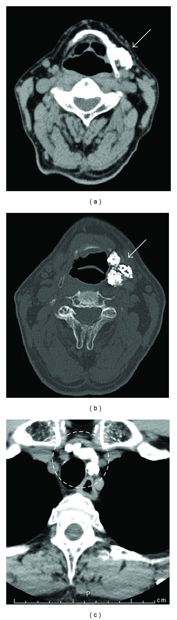
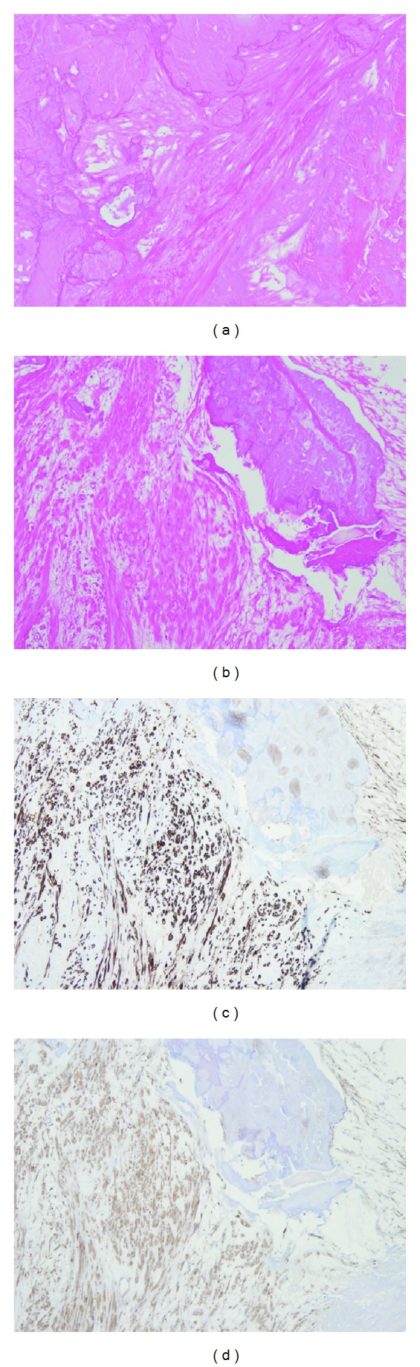
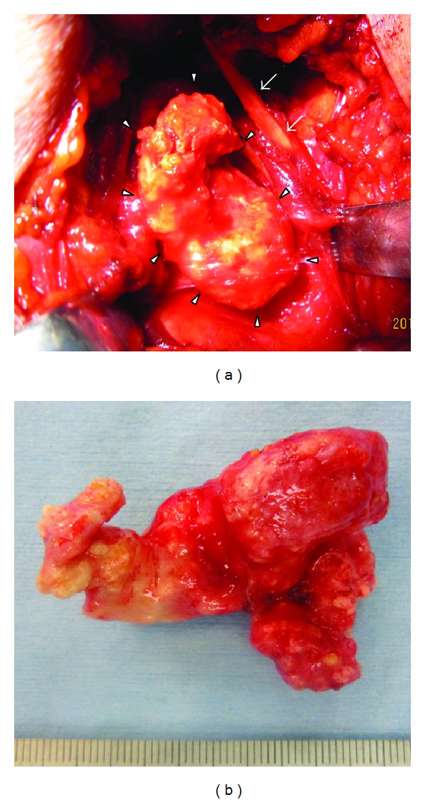
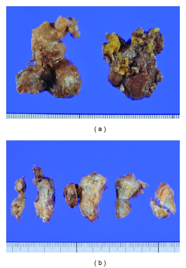
We encountered a patient with primary cervical leiomyoma with remarkable calcification and ossification. A 68-year-old man presenting with induration and swelling of the left submandibular region was found to have nodular lesions with calcifications in the left submandibular region and the upper mediastinum on CT. Fine needle aspiration biopsies (FNAB) of the left submandibular lesion revealed no malignancy. Resection was performed for definitive diagnosis and treatment. The resected specimen contained a solid tumor, which was markedly calcified and ossified on the cut surface. Histopathological examination showed proliferating spindle cells in a tangled and crossed arrangement. Immunohistochemically, the spindle cells were stained intensely with α-SMA and h-caldesmon, consistent with smooth muscle cells. These findings led to a definitive diagnosis of leiomyoma with calcification and ossification. This is extremely rare and the preoperative differentiation from other tumors of the head and neck was very difficult. By resection of the submandibular tumor, both definitive diagnosis of leiomyoma by histopathological and immunohistochemical analyses and treatment could be carried out. However, as the tumor in the upper mediastinum was most likely to be leiomyoma with calcification, he did not wish to undergo its biopsy and resection immediately. We have continued the follow-up.
我们遇到了一名患有原发性宫颈平滑肌瘤且伴有显著钙化和骨化的患者。一名68岁男性,因左下颌下区硬结和肿胀就诊,CT检查发现左下颌下区及上纵隔有结节状钙化病变。左下颌下病变的细针穿刺活检(FNAB)未发现恶性肿瘤。为明确诊断和治疗进行了切除手术。切除标本为实性肿瘤,切面可见明显钙化和骨化。组织病理学检查显示梭形细胞呈交织状增生排列。免疫组化显示,梭形细胞α-SMA和h-钙调蛋白染色强烈,符合平滑肌细胞特征。这些结果明确诊断为伴有钙化和骨化的平滑肌瘤。这种情况极为罕见,术前与头颈部其他肿瘤进行鉴别诊断非常困难。通过切除下颌下肿瘤,既可以通过组织病理学和免疫组化分析明确平滑肌瘤诊断,又能进行治疗。然而,由于上纵隔的肿瘤很可能是伴有钙化的平滑肌瘤,他不希望立即进行活检和切除。我们一直在进行随访。