Collister John P, Nahey David B, Hendel Michael D, Brooks Virginia L
Department of Veterinary and Biomedical Sciences, College of Veterinary Medicine, University of Minnesota, St. Paul, 55108, Minnesota.
Department of Physiology & Pharmacology, Oregon Health and Science University Portland, Oregon, 97239.
Physiol Rep. 2014 Jan 6;2(1):e00191. doi: 10.1002/phy2.191. eCollection 2014 Jan 1.
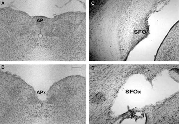
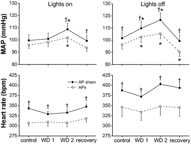
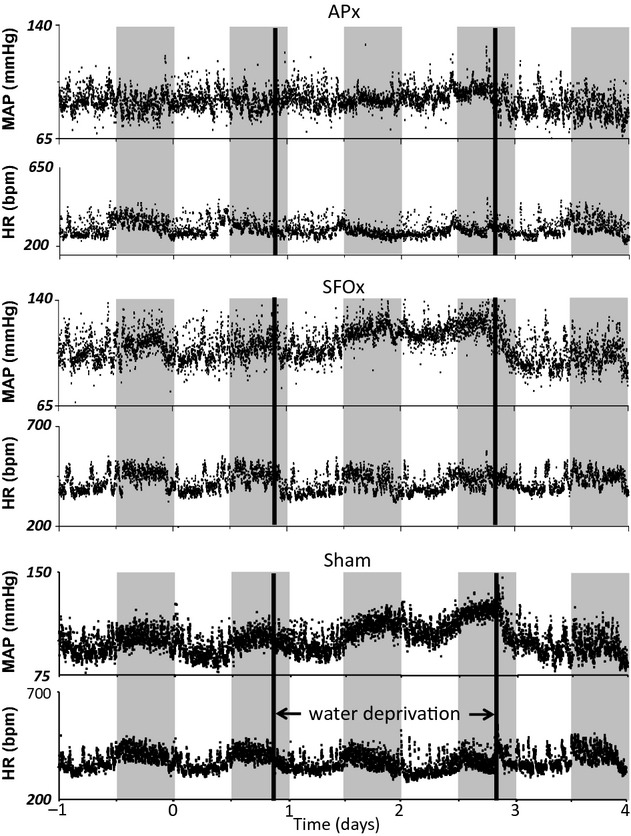
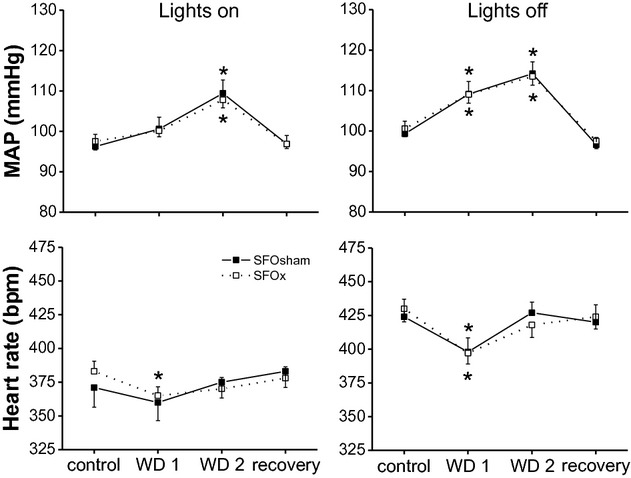
In rats, water deprivation (WD) increases arterial blood pressure (BP) in part due to actions of elevated osmolality in the brain to increase vasopressin levels and sympathetic activity. However, the osmoreceptors that mediate this response have not been identified. To test the hypothesis that osmoregulatory circumventricular organs are involved, BP and heart rate (HR) were continuously recorded telemetrically during 48 h of WD in normal rats with lesions (x) or sham lesions (sham) of the subfornical organ (SFO) or area postrema (AP). Although WD increased BP in SFOx and SFOsham rats, no significant difference in the hypertensive response was observed between groups. HR decreased transiently but similarly in SFOx and SFOsham rats during the first 24 h of WD. When water was reintroduced, BP and HR decreased rapidly and similarly in both groups. BP (during lights off) and HR were both lower in APx rats before WD compared to APsham. WD increased BP less in APx rats, and the transient bradycardia was eliminated. Upon reintroduction of drinking water, smaller falls in both BP and HR were observed in APx rats compared to APsham rats. WD increased plasma osmolality and vasopressin levels similarly in APx and APsham rats, and acute blockade of systemic V1 vasopressin receptors elicited similar depressor responses, suggesting that the attenuated BP response is not due to smaller increases in vasopressin or osmolality. In conclusion, the AP, but not the SFO, is required for the maximal hypertensive effect induced by WD in rats.
在大鼠中,缺水(WD)会使动脉血压(BP)升高,部分原因是大脑中渗透压升高会增加血管加压素水平和交感神经活动。然而,介导这种反应的渗透压感受器尚未被确定。为了检验渗透调节室周器官参与其中这一假设,在对穹窿下器官(SFO)或最后区(AP)进行损伤(x)或假损伤(假手术)的正常大鼠缺水48小时期间,通过遥测连续记录血压(BP)和心率(HR)。尽管缺水使SFOx和SFO假手术大鼠的血压升高,但两组之间在高血压反应方面未观察到显著差异。在缺水的前24小时内,SFOx和SFO假手术大鼠的心率均短暂下降,但下降情况相似。当重新给予水时,两组的血压和心率均迅速下降且下降情况相似。与AP假手术大鼠相比,APx大鼠在缺水前(熄灯期间)的血压和心率均较低。APx大鼠缺水时血压升高幅度较小,且短暂的心动过缓消失。重新给予饮用水后,与AP假手术大鼠相比,APx大鼠的血压和心率下降幅度较小。APx和AP假手术大鼠缺水时血浆渗透压和血管加压素水平升高情况相似,急性阻断全身V1血管加压素受体引发的降压反应相似,这表明血压反应减弱并非由于血管加压素或渗透压升高幅度较小所致。总之,大鼠缺水诱导的最大高血压效应需要AP参与,但不需要SFO参与。