Zhang Jiao, Ho Jenny Chung-Yee, Chan Yau-Chi, Lian Qizhou, Siu Chung-Wah, Tse Hung-Fat
Division of Cardiology, Department of Medicine, Queen Mary Hospital, The University of Hong Kong, Hong Kong SAR, China.
Division of Cardiology, Department of Medicine, Queen Mary Hospital, The University of Hong Kong, Hong Kong SAR, China ; Research Centre of Heart, Brain, Hormone & Healthy Aging, Li Ka Shing Faculty of Medicine, The University of Hong Kong, Hong Kong SAR, China.
Physiol Rep. 2014 Feb 25;2(2):e00237. doi: 10.1002/phy2.237. eCollection 2014 Feb 1.
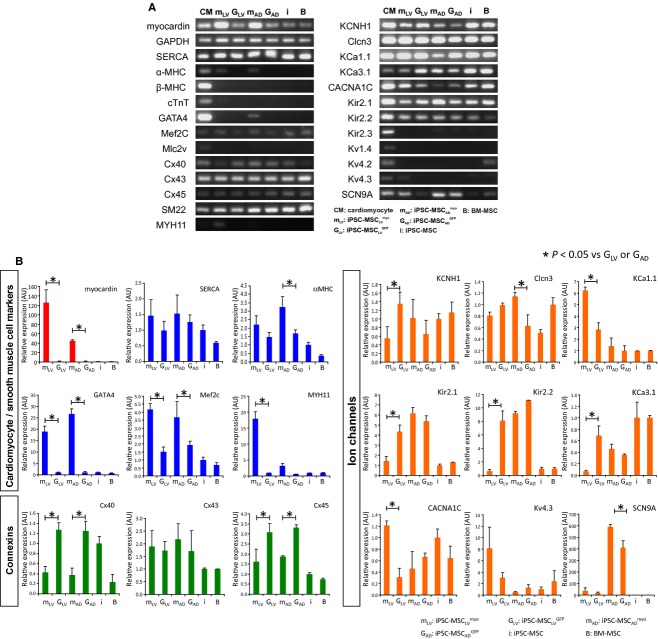
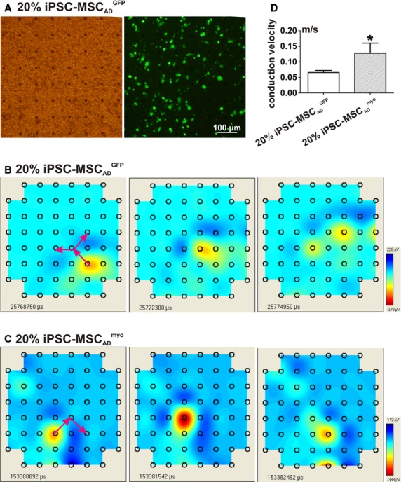
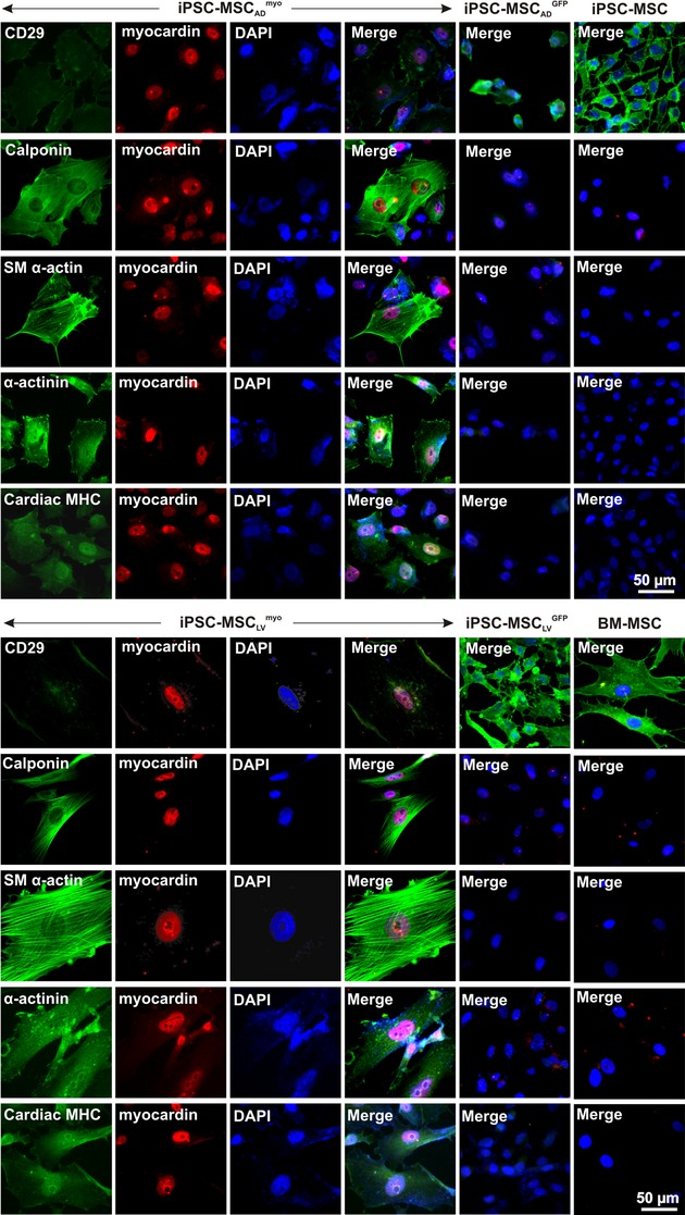
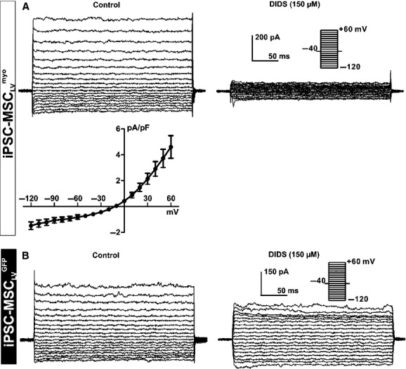
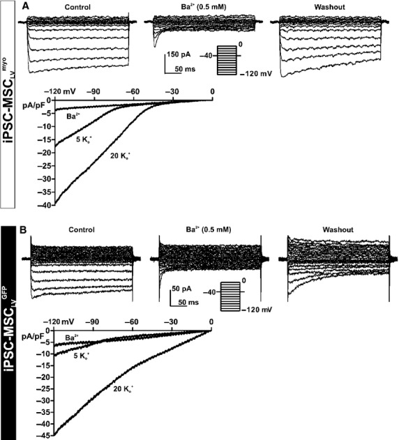
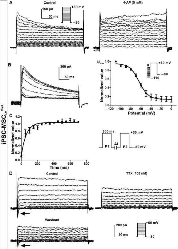
Mesenchymal stem cells (MSCs) derived from human-induced pluripotent stem cells (iPSCs) show superior proliferative capacity and therapeutic potential than those derived from bone marrow (BM). Ectopic expression of myocardin further improved the therapeutic potential of BM-MSCs in a mouse model of myocardial infarction. The aim was of this study was to assess whether forced myocardin expression in iPSC-MSCs could further enhance their transdifferentiation to cardiomyocytes and improve their electrophysiological properties for cardiac regeneration. Myocardin was overexpressed in iPSC-MSCs using viral vectors (adenovirus or lentivirus). The expression of smooth muscle cell and cardiomyocyte markers, and ion channel genes was examined by reverse transcription-polymerase chain reaction (RT-PCR), immunofluorescence staining and patch clamp. The conduction velocity of the neonatal rat ventricular cardiomyocytes cocultured with iPSC-MSC monolayer was measured by multielectrode arrays recording plate. Myocardin induced the expression of α-MHC, GATA4, α-actinin, cardiac MHC, MYH11, calponin, and SM α-actin, but not cTnT, β-MHC, and MLC2v in iPSC-MSCs. Overexpression of myocardin in iPSC-MSC enhanced the expression of SCN9A and CACNA1C, but reduced that of KCa3.1 and Kir2.2 in iPSC-MSCs. Moreover, BKCa, IKir, ICl, Ito and INa.TTX were detected in iPSC-MSC with myocardin overexpression; while only BKCa, IKir, ICl, IKDR, and IKCa were noted in iPSC-MSC transfected with green florescence protein. Furthermore, the conduction velocity of iPSC-MSC was significantly increased after myocardin overexpression. Overexpression of myocardin in iPSC-MSCs resulted in partial transdifferentiation into cardiomyocytes phenotype and improved the electrical conduction during integration with mature cardiomyocytes.
源自人诱导多能干细胞(iPSC)的间充质干细胞(MSC)比源自骨髓(BM)的间充质干细胞具有更强的增殖能力和治疗潜力。在心肌梗死小鼠模型中,心肌转录因子的异位表达进一步提高了BM-MSC的治疗潜力。本研究旨在评估在iPSC-MSC中强制表达心肌转录因子是否能进一步增强其向心肌细胞的转分化能力,并改善其心脏再生的电生理特性。使用病毒载体(腺病毒或慢病毒)在iPSC-MSC中过表达心肌转录因子。通过逆转录聚合酶链反应(RT-PCR)、免疫荧光染色和膜片钳技术检测平滑肌细胞和心肌细胞标志物以及离子通道基因的表达。通过多电极阵列记录板测量与iPSC-MSC单层共培养的新生大鼠心室心肌细胞的传导速度。心肌转录因子诱导了iPSC-MSC中α-MHC、GATA4、α-肌动蛋白、心肌MHC、MYH11、钙调蛋白和SMα-肌动蛋白的表达,但未诱导cTnT、β-MHC和MLC2v的表达。iPSC-MSC中过表达心肌转录因子增强了SCN9A和CACNA1C的表达,但降低了KCa3.1和Kir2.2的表达。此外,在过表达心肌转录因子的iPSC-MSC中检测到BKCa、IKir、ICl、Ito和INa.TTX;而在转染绿色荧光蛋白的iPSC-MSC中仅检测到BKCa、IKir、ICl、IKDR和IKCa。此外,过表达心肌转录因子后iPSC-MSC的传导速度显著增加。iPSC-MSC中过表达心肌转录因子导致部分转分化为心肌细胞表型,并改善了与成熟心肌细胞整合过程中的电传导。