Mazzotta Cosimo, Balestrazzi Angelo, Traversi Claudio, Caragiuli Stefano, Caporossi Aldo
Department of Ophthalmology, Siena University Hospital, Siena, Italy.
Department of Ophthalmology, Rome Catholic University, Rome, Italy.
Case Rep Ophthalmol. 2014 Apr 12;5(1):125-31. doi: 10.1159/000362327. eCollection 2014 Jan.
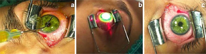
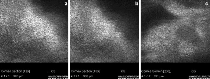
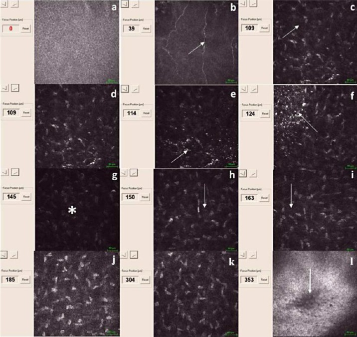
We report the first pilot qualitative confocal microscopic analysis of a laser in situ keratomileusis (Lasik) treatment combined with sequential high-fluence accelerated corneal collagen cross-linking, denominated Lasik XTra, by means of HRT II laser scanning in vivo confocal microscopy after a 6-month follow-up. After obtaining approval from the Siena University Hospital Institutional Review Board, a 33-year-old female patient underwent a Lasik XTra procedure in her left eye. Confocal analysis demonstrated induced slight corneal microstructural changes by the interaction between UV-A, riboflavin and corneal stromal collagen, beyond the interface to a depth of 160 µm, without adverse events at the interface and endothelial levels. This application may be considered a prophylactic biomechanical treatment, stiffening the intermediate corneal stroma to prevent corneal ectasia and stabilizing the clinical results of refractive surgery. According to our preliminary experiences, this combined approach may be useful in higher-risk Lasik patients for hyperopic treatments, high myopia and lower corneal thicknesses.
我们报告了首例对激光原位角膜磨镶术(Lasik)联合序贯高能量加速角膜胶原交联(命名为Lasik XTra)进行的定性共焦显微镜分析试点研究,该研究通过HRT II激光扫描活体共焦显微镜在6个月随访后进行。在获得锡耶纳大学医院机构审查委员会的批准后,一名33岁女性患者左眼接受了Lasik XTra手术。共焦分析表明,紫外线A、核黄素与角膜基质胶原之间的相互作用在角膜界面以外160 µm深度处引起了轻微的角膜微观结构变化,在界面和内皮层面未出现不良事件。这种应用可被视为一种预防性生物力学治疗,强化角膜中间基质以预防角膜扩张,并稳定屈光手术的临床效果。根据我们的初步经验,这种联合方法可能对远视治疗、高度近视和角膜厚度较低的高风险Lasik患者有用。