Yamada Takashi, Fedotovskaya Olga, Cheng Arthur J, Cornachione Anabelle S, Minozzo Fabio C, Aulin Cecilia, Fridén Cecilia, Turesson Carl, Andersson Daniel C, Glenmark Birgitta, Lundberg Ingrid E, Rassier Dilson E, Westerblad Håkan, Lanner Johanna T
Department of Physiology and Pharmacology, Karolinska Institutet, Stockholm, Sweden School of Health Sciences, Sapporo Medical University, Sapporo, Japan.
Department of Physiology and Pharmacology, Karolinska Institutet, Stockholm, Sweden.
Ann Rheum Dis. 2015 Oct;74(10):1907-14. doi: 10.1136/annrheumdis-2013-205007. Epub 2014 May 22.
Skeletal muscle weakness is a prominent clinical feature in patients with rheumatoid arthritis (RA), but the underlying mechanism(s) is unknown. Here we investigate the mechanisms behind arthritis-induced skeletal muscle weakness with special focus on the role of nitrosative stress on intracellular Ca(2+) handling and specific force production.
Nitric oxide synthase (NOS) expression, degree of nitrosative stress and composition of the major intracellular Ca(2+) release channel (ryanodine receptor 1, RyR1) complex were measured in muscle. Changes in cytosolic free Ca(2+) concentration ([Ca(2+)]i) and force production were assessed in single-muscle fibres and isolated myofibrils using atomic force cantilevers.
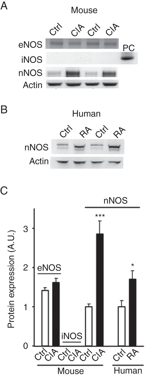
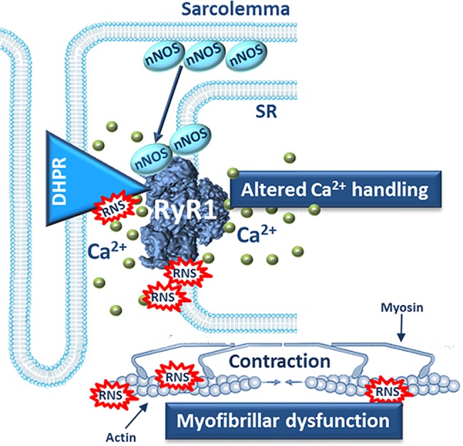
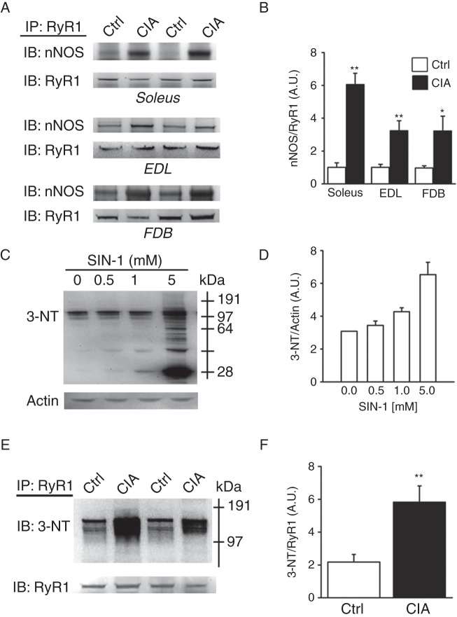
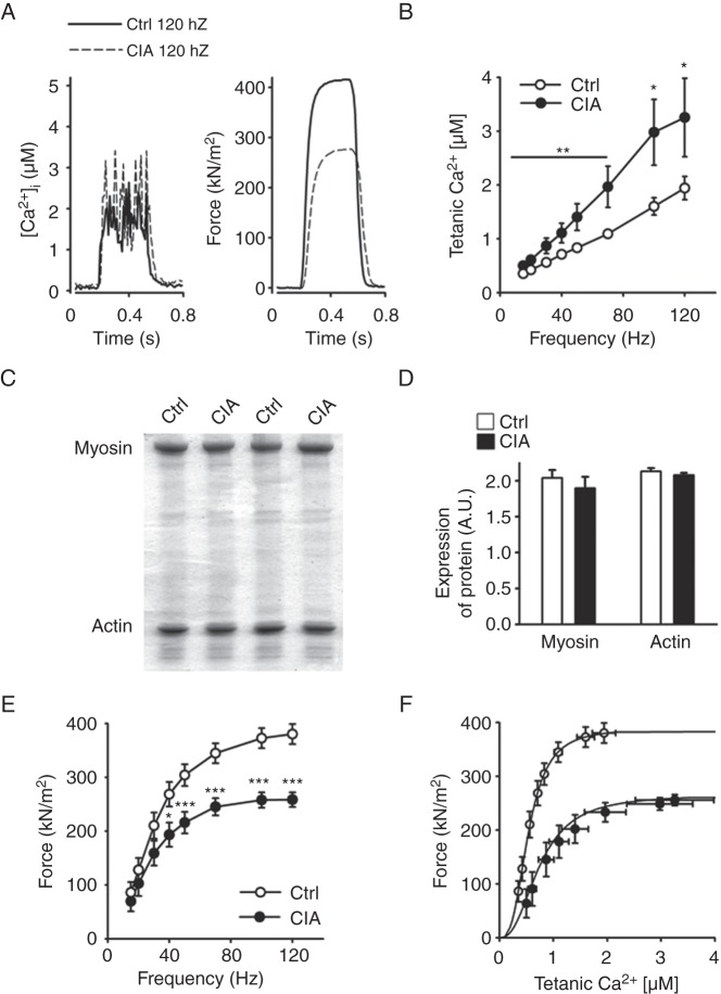
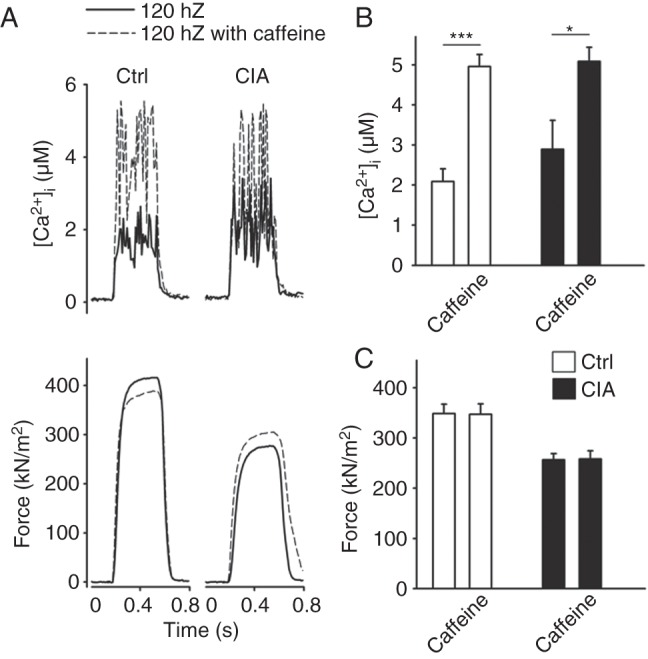
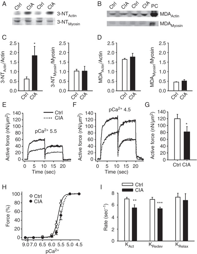
The total neuronal NOS (nNOS) levels were increased in muscles both from collagen-induced arthritis (CIA) mice and patients with RA. The nNOS associated with RyR1 was increased and accompanied by increased [Ca(2+)]i during contractions of muscles from CIA mice. A marker of peroxynitrite-derived nitrosative stress (3-nitrotyrosine, 3-NT) was increased on the RyR1 complex and on actin of muscles from CIA mice. Despite increased [Ca(2+)]i, individual CIA muscle fibres were weaker than in healthy controls, that is, force per cross-sectional area was decreased. Furthermore, force and kinetics were impaired in CIA myofibrils, hence actin and myosin showed decreased ability to interact, which could be a result of increased 3-NT content on actin.
Arthritis-induced muscle weakness is linked to nitrosative modifications of the RyR1 protein complex and actin, which are driven by increased nNOS associated with RyR1 and progressively increasing Ca(2+) activation.
骨骼肌无力是类风湿关节炎(RA)患者的一个突出临床特征,但其潜在机制尚不清楚。在此,我们研究关节炎诱导的骨骼肌无力背后的机制,特别关注亚硝化应激在细胞内钙(Ca2+)处理和比肌力产生中的作用。
检测肌肉中一氧化氮合酶(NOS)的表达、亚硝化应激程度以及主要细胞内钙释放通道(兰尼碱受体1,RyR1)复合物的组成。使用原子力悬臂在单根肌纤维和分离的肌原纤维中评估细胞溶质游离钙浓度([Ca2+]i)和肌力产生的变化。
胶原诱导性关节炎(CIA)小鼠和RA患者肌肉中的总神经元型NOS(nNOS)水平均升高。在CIA小鼠肌肉收缩过程中,与RyR1相关的nNOS增加,并伴有[Ca2+]i升高。CIA小鼠肌肉的RyR1复合物和肌动蛋白上的过氧亚硝酸盐衍生的亚硝化应激标志物(3-硝基酪氨酸,3-NT)增加。尽管[Ca2+]i升高,但单个CIA肌纤维比健康对照中的肌纤维更弱,即每横截面积的力量降低。此外,CIA肌原纤维的力量和动力学受损,因此肌动蛋白和肌球蛋白的相互作用能力下降,这可能是肌动蛋白上3-NT含量增加的结果。
关节炎诱导的肌肉无力与RyR1蛋白复合物和肌动蛋白的亚硝化修饰有关,这是由与RyR1相关的nNOS增加和Ca2+激活逐渐增加所驱动的。