• 文献检索
  • 文档翻译
  • 深度研究
  • 学术资讯
  • Suppr Zotero 插件Zotero 插件
  • 邀请有礼
  • 套餐&价格
  • 历史记录
应用&插件
Suppr Zotero 插件Zotero 插件浏览器插件Mac 客户端Windows 客户端微信小程序
定价
高级版会员购买积分包购买API积分包
服务
文献检索文档翻译深度研究API 文档MCP 服务
关于我们
关于 Suppr公司介绍联系我们用户协议隐私条款
关注我们

Suppr 超能文献

核心技术专利:CN118964589B侵权必究
粤ICP备2023148730 号-1Suppr @ 2026

文献检索

告别复杂PubMed语法,用中文像聊天一样搜索,搜遍4000万医学文献。AI智能推荐,让科研检索更轻松。

立即免费搜索

文件翻译

保留排版,准确专业,支持PDF/Word/PPT等文件格式,支持 12+语言互译。

免费翻译文档

深度研究

AI帮你快速写综述,25分钟生成高质量综述,智能提取关键信息,辅助科研写作。

立即免费体验

[多层螺旋CT对周围型肺癌伴干性胸膜播散的影像诊断]

[MSCT imaging diagnosis of peripheral lung cancer with dry pleural dissemination].

作者信息

Liu Jia, Li Wenwu, Huang Yong, Liu Yuhui

机构信息

Department of Radiology, Shandong Tumor Hospital, Affilialed to Shandong Acadaemy of Medical Sciences, Jinan 250017, China;School of Medicine and Life Scineces, University of Jinan-Shandong Acadamy of Medical Scineces, Jinan 250007, China.

Department of Radiology, Shandong Tumor Hospital, Affilialed to Shandong Acadaemy of Medical Sciences, Jinan 250017, China.

出版信息

Zhongguo Fei Ai Za Zhi. 2014 May;17(5):406-10. doi: 10.3779/j.issn.1009-3419.2014.05.07.

DOI:10.3779/j.issn.1009-3419.2014.05.07
PMID:24854558
原文链接:https://pmc.ncbi.nlm.nih.gov/articles/PMC6000440/
Abstract

BACKGROUND AND OBJECTIVE

The rate of pleural metastasis in peripheral lung cancer is high, and the dry pleural metastasis easily missed diagnosis preoperatively and cause unnecessary surgery. Therefore, preoperative diagnosis is particularly important. To review the multislice spiral computed tomography (MSCT) image of peripheral lung cancer with dry pleural dissemination, and to discuss its diagnostic value for understanding the dry pleural dissemination.

METHODS

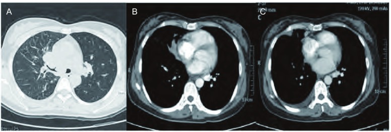
Reviewed and analyzed the MSCT images of pathologically or clinically diagnosed peripheral lung cancer with dry pleural dissemination in 27 patients. Analyze the imaging characteristics respectively from pleural thickening and pleural nodules.

RESULTS

The dry pleural dissemination of lung cancer were detected in 85% by CT. The rate of CT detection of dissemination on the visceral pleura and the interlobar pleura were 63% and 91%, respectively. 26 cases were with multiple pleural nodules, all were located on the same side with the primary lesions; 8 cases were with peritoneum visceralis nodules that are mostly circular with diameters of 3 mm-15 mm. The lung-nodules interfaces were clear. 23 cases had interlobar pleura nodules (all with more than 6 nodules), some are big (diameter >5 mm) while some are small (diameter <5 mm). The nodules are arranged along the interlobar pleura as beaded string or in clusters around the interlobar pleura. 15 cases were with pleural thickening, including band-like, uneven, or both exist at the same time. There are more mixed type in this group (63%).

CONCLUSIONS

MSCT has great diagnostic value for peripheral lung cancer with dry pleural dissemination, especially with high accuracy of pleural nodules.

摘要

背景与目的

周围型肺癌胸膜转移率较高,干性胸膜转移术前易漏诊,导致不必要的手术。因此,术前诊断尤为重要。回顾分析周围型肺癌干性胸膜播散的多层螺旋计算机断层扫描(MSCT)图像,探讨其对干性胸膜播散的诊断价值。

方法

回顾性分析27例经病理或临床诊断为周围型肺癌干性胸膜播散的MSCT图像。分别从胸膜增厚和胸膜结节方面分析其影像特征。

结果

CT对肺癌干性胸膜播散的检出率为85%。脏层胸膜和叶间胸膜播散的CT检出率分别为63%和91%。26例有多个胸膜结节,均位于原发灶同侧;8例有脏层胸膜结节,多呈圆形,直径3mm - 15mm,肺结节界面清晰。23例有叶间胸膜结节(均超过6个),部分较大(直径>5mm),部分较小(直径<5mm),结节沿叶间胸膜呈串珠状排列或在叶间胸膜周围簇状分布。15例有胸膜增厚,包括带状、不均匀或两者同时存在,本组以混合型居多(63%)。

结论

MSCT对周围型肺癌干性胸膜播散具有较大诊断价值,尤其是对胸膜结节的诊断准确性较高。

https://cdn.ncbi.nlm.nih.gov/pmc/blobs/e3fc/6000440/4bfc866f3cef/zgfazz-17-5-406-4.jpg
https://cdn.ncbi.nlm.nih.gov/pmc/blobs/e3fc/6000440/33e530a64e5f/zgfazz-17-5-406-1.jpg
https://cdn.ncbi.nlm.nih.gov/pmc/blobs/e3fc/6000440/f7839d1e7145/zgfazz-17-5-406-2.jpg
https://cdn.ncbi.nlm.nih.gov/pmc/blobs/e3fc/6000440/82d84c39bee9/zgfazz-17-5-406-3.jpg
https://cdn.ncbi.nlm.nih.gov/pmc/blobs/e3fc/6000440/4bfc866f3cef/zgfazz-17-5-406-4.jpg

相似文献

1
[MSCT imaging diagnosis of peripheral lung cancer with dry pleural dissemination].[多层螺旋CT对周围型肺癌伴干性胸膜播散的影像诊断]
Zhongguo Fei Ai Za Zhi. 2014 May;17(5):406-10. doi: 10.3779/j.issn.1009-3419.2014.05.07.
2
Integrated PET/CT and the dry pleural dissemination of peripheral adenocarcinoma of the lung: diagnostic implications.PET/CT融合成像与肺外周腺癌的干性胸膜播散:诊断意义
J Comput Assist Tomogr. 2006 Jan-Feb;30(1):70-6. doi: 10.1097/01.rct.0000185383.50636.d1.
3
[Diagnostic value of multislice spiral CT and MRI in detection of tumor recurrence after liver transplantation for hepatocellular carcinoma].多层螺旋CT与MRI对肝细胞癌肝移植术后肿瘤复发的诊断价值
Zhonghua Zhong Liu Za Zhi. 2009 Sep;31(9):691-6.
4
CT diagnosis of pleural dissemination without pleural effusion in primary lung cancer.原发性肺癌无胸腔积液的胸膜播散的CT诊断
Radiat Med. 1996 May-Jun;14(3):117-9.
5
Dry pleural dissemination in non-small cell lung cancer: prognostic and diagnostic implications.非小细胞肺癌的干性胸膜播散:预后和诊断意义。
Radiology. 2011 Aug;260(2):568-74. doi: 10.1148/radiol.11110053. Epub 2011 Jun 3.
6
Pleural dissemination in non-small cell lung cancer: results of radiological evaluation and surgical treatment.
J Surg Oncol. 1994 Sep;57(1):33-9. doi: 10.1002/jso.2930570111.
7
Dry small pleural dissemination of adenocarcinoma of the lung preoperatively detected by PET/CT: a report of two cases.PET/CT术前检测到的肺腺癌干性小胸膜播散:2例报告
Acta Med Okayama. 2008 Feb;62(1):55-8. doi: 10.18926/AMO/30991.
8
[CT features and anatomic distribution of pleural dissemination in lung cancer].[肺癌胸膜播散的CT特征及解剖分布]
Zhongguo Fei Ai Za Zhi. 2003 Feb 20;6(1):26-9. doi: 10.3779/j.issn.1009-3419.2003.01.07.
9
Efficacy of Multi-slice Spiral CT and Rapid On-site Evaluation in Diagnosis of Pulmonary Nodules.多层螺旋 CT 及现场快速评估在肺结节诊断中的效能。
Altern Ther Health Med. 2024 Jul;30(7):222-227.
10
Multidetector CT Findings and Differential Diagnoses of Malignant Pleural Mesothelioma and Metastatic Pleural Diseases in Korea.韩国恶性胸膜间皮瘤及胸膜转移瘤的多排螺旋CT表现与鉴别诊断
Korean J Radiol. 2016 Jul-Aug;17(4):545-53. doi: 10.3348/kjr.2016.17.4.545. Epub 2016 Jun 27.

引用本文的文献

1
[Analysis of Influencing Factors of Complications for CT-guided 
Percutaneous Lung Biopsy].[CT引导下经皮肺穿刺活检并发症的影响因素分析]
Zhongguo Fei Ai Za Zhi. 2024 Mar 20;27(3):179-186. doi: 10.3779/j.issn.1009-3419.2024.101.03.

本文引用的文献

1
Thin-section CT as an optimal diagnostic tool in the evaluation of dry pleural dissemination in non-small cell lung cancer.
Radiology. 2012 Jan;262(1):368-9; author reply 369. doi: 10.1148/radiol.11111547.
2
Dry pleural dissemination in non-small cell lung cancer: prognostic and diagnostic implications.非小细胞肺癌的干性胸膜播散:预后和诊断意义。
Radiology. 2011 Aug;260(2):568-74. doi: 10.1148/radiol.11110053. Epub 2011 Jun 3.
3
The new lung cancer staging system.新的肺癌分期系统。
Chest. 2009 Jul;136(1):260-271. doi: 10.1378/chest.08-0978.
4
Dry small pleural dissemination of adenocarcinoma of the lung preoperatively detected by PET/CT: a report of two cases.PET/CT术前检测到的肺腺癌干性小胸膜播散:2例报告
Acta Med Okayama. 2008 Feb;62(1):55-8. doi: 10.18926/AMO/30991.
5
Integrated PET/CT and the dry pleural dissemination of peripheral adenocarcinoma of the lung: diagnostic implications.PET/CT融合成像与肺外周腺癌的干性胸膜播散:诊断意义
J Comput Assist Tomogr. 2006 Jan-Feb;30(1):70-6. doi: 10.1097/01.rct.0000185383.50636.d1.
6
Retrospective review of lung cancer patients with pleural dissemination after limited operations combined with parietal pleurectomy.对接受有限手术联合壁层胸膜切除术治疗后发生胸膜播散的肺癌患者进行回顾性分析。
J Surg Oncol. 2005 Sep 15;91(4):237-42. doi: 10.1002/jso.20333.
7
Helical computed tomography diagnosis of pleural dissemination in lung cancer: comparison of thick-section and thin-section helical computed tomography.螺旋计算机断层扫描对肺癌胸膜播散的诊断:厚层与薄层螺旋计算机断层扫描的比较
J Thorac Imaging. 1998 Jul;13(3):211-8. doi: 10.1097/00005382-199807000-00009.
8
CT diagnosis of pleural dissemination without pleural effusion in primary lung cancer.原发性肺癌无胸腔积液的胸膜播散的CT诊断
Radiat Med. 1996 May-Jun;14(3):117-9.
9
Prevalence of pathologically proven intrapulmonary lymph nodes and their appearance on CT.
AJR Am J Roentgenol. 1996 Sep;167(3):629-30. doi: 10.2214/ajr.167.3.8751667.