Reeves Stephen R, Kolstad Tessa, Lien Tin-Yu, Elliott Molly, Ziegler Steven F, Wight Thomas N, Debley Jason S
Division of Pulmonary Medicine, Seattle Children's Hospital, University of Washington, Seattle, Wash; Center for Immunity and Immunotherapies, Seattle Children's Research Institute, Seattle, Wash.
Center for Immunity and Immunotherapies, Seattle Children's Research Institute, Seattle, Wash.
J Allergy Clin Immunol. 2014 Sep;134(3):663-670.e1. doi: 10.1016/j.jaci.2014.04.007. Epub 2014 May 27.
Airway remodeling might explain lung function decline among asthmatic children. Extracellular matrix (ECM) deposition by human lung fibroblasts (HLFs) is implicated in airway remodeling. Airway epithelial cell (AEC) signaling might regulate HLF ECM expression.
We sought to determine whether AECs from asthmatic children differentially regulate HLF expression of ECM constituents.
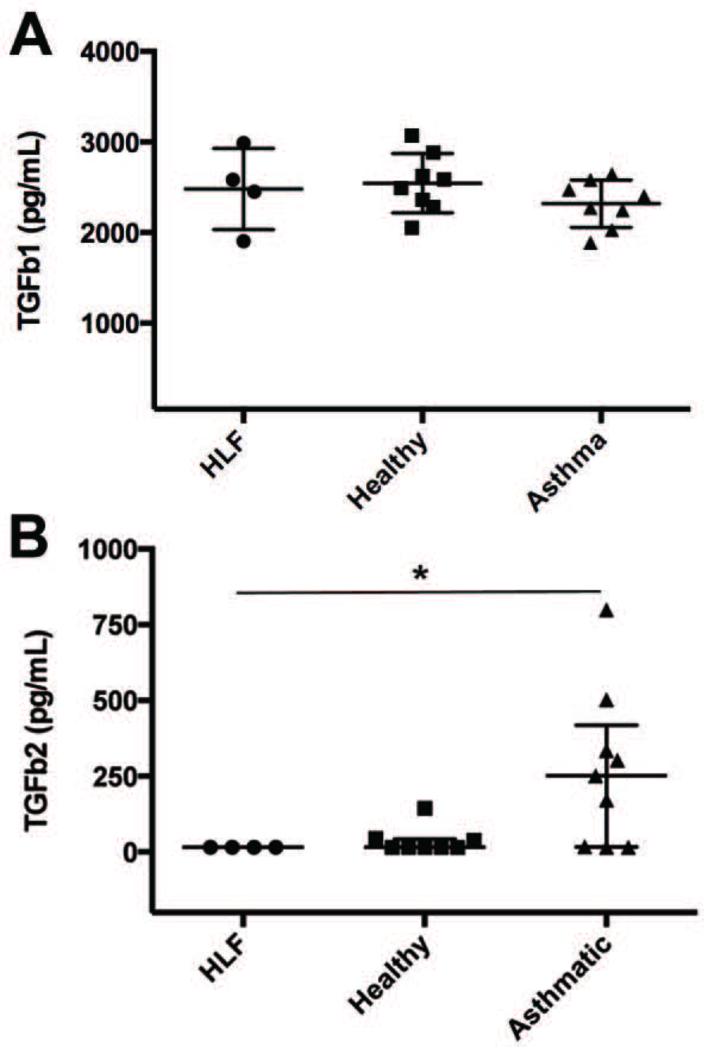
Primary AECs were obtained from well-characterized atopic asthmatic (n = 10) and healthy (n = 10) children intubated during anesthesia for an elective surgical procedure. AECs were differentiated at an air-liquid interface for 3 weeks and then cocultured with HLFs from a healthy child for 96 hours. Collagen I (COL1A1), collagen III (COL3A1), hyaluronan synthase (HAS) 2, and fibronectin expression by HLFs and prostaglandin E2 synthase (PGE2S) expression by AECs were assessed by using RT-PCR. TGF-β1 and TGF-β2 concentrations in media were measured by using ELISA.
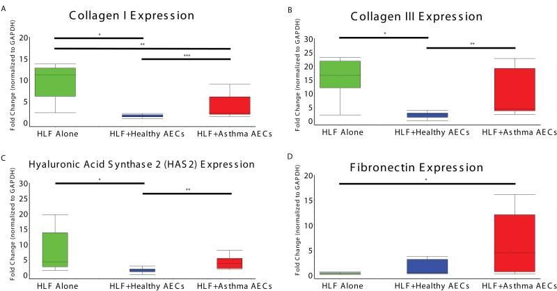
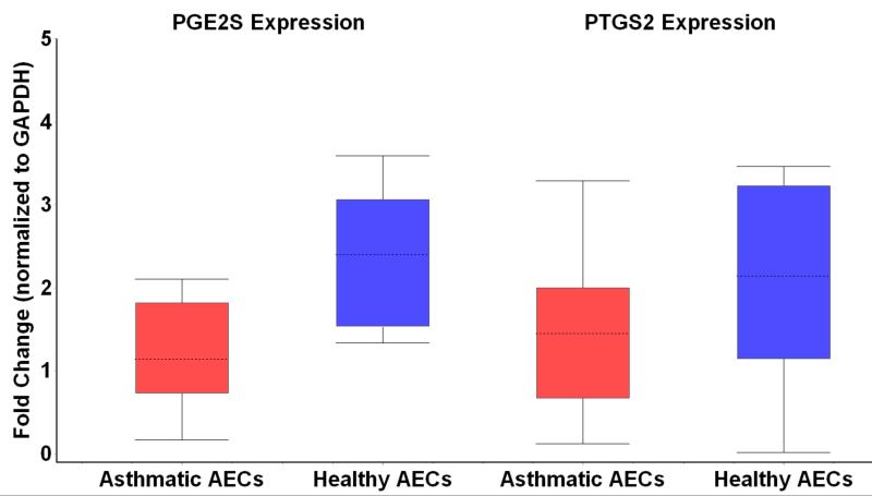
COL1A1 and COL3A1 expression by HLFs cocultured with AECs from asthmatic patients was greater than that by HLFs cocultured with AECs from healthy subjects (2.2-fold, P < .02; 10.8-fold, P < .02). HAS2 expression by HLFs cocultured with AECs from asthmatic patients was 2.5-fold higher than that by HLFs cocultured with AECs from healthy subjects (P < .002). Fibronectin expression by HLFs cocultured with AECs from asthmatic patients was significantly greater than that by HLFs alone. TGF-β2 activity was increased in cocultures of HLFs with AECs from asthmatic patients (P < .05), whereas PGES2 was downregulated in AEC-HLF cocultures (2.2-fold, P < .006).
HLFs cocultured with AECs from asthmatic patients showed differential expression of the ECM constituents COL1A1 and COL3A1 and HAS2 compared with HLFs cocultured with AECs from healthy subjects. These findings support a role for altered ECM production in asthmatic airway remodeling, possibly regulated by unbalanced AEC signaling.
气道重塑可能解释哮喘儿童肺功能下降的原因。人肺成纤维细胞(HLF)的细胞外基质(ECM)沉积与气道重塑有关。气道上皮细胞(AEC)信号可能调节HLF的ECM表达。
我们试图确定哮喘儿童的AEC是否对HLF的ECM成分表达有不同的调节作用。
从在择期外科手术麻醉期间插管的明确诊断为特应性哮喘的儿童(n = 10)和健康儿童(n = 10)中获取原代AEC。将AEC在气液界面分化3周,然后与一名健康儿童的HLF共培养96小时。通过逆转录聚合酶链反应(RT-PCR)评估HLF的I型胶原(COL1A1)、III型胶原(COL3A1)、透明质酸合酶(HAS)2和纤连蛋白的表达以及AEC的前列腺素E2合酶(PGE2S)的表达。使用酶联免疫吸附测定(ELISA)测量培养基中转化生长因子-β1(TGF-β1)和转化生长因子-β2(TGF-β2)的浓度。
与来自健康受试者的AEC共培养的HLF相比,与哮喘患者的AEC共培养的HLF的COL1A1和COL3A1表达更高(分别为2.2倍,P < 0.02;10.8倍,P < 0.02)。与来自哮喘患者的AEC共培养的HLF的HAS2表达比与来自健康受试者的AEC共培养的HLF高2.5倍(P < 0.002)。与哮喘患者的AEC共培养的HLF的纤连蛋白表达明显高于单独培养的HLF。在与哮喘患者的AEC共培养的HLF中,TGF-β2活性增加(P < 0.05),而在AEC-HLF共培养物中前列腺素E2合酶(PGES2)下调(2.2倍,P < 0.006)。
与来自健康受试者的AEC共培养的HLF相比,与哮喘患者的AEC共培养的HLF显示出ECM成分COL1A1、COL3A1和HAS2的差异表达。这些发现支持ECM产生改变在哮喘气道重塑中的作用,可能由不平衡的AEC信号调节。