Aslam Muhammad I, Flexer Susannah M, Figueiredo Rodrego, Ashour Hamdy Y, Bhattacharya Vish
Department of General Surgery, Ayr University Hospital, Ayrshire and Arran NHS Trust, South Ayrshire KA6 6DX, UK.
Department of General Surgery, Ayr University Hospital, Ayrshire and Arran NHS Trust, South Ayrshire KA6 6DX, UK
J Surg Case Rep. 2014 Feb 6;2014(2). doi: 10.1093/jscr/rjt129. Print 2014 Feb.
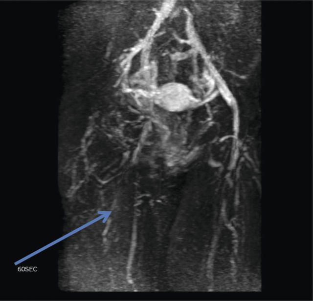
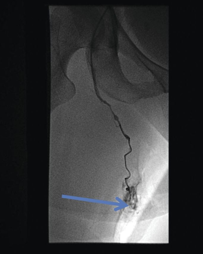
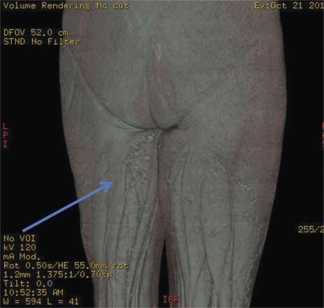
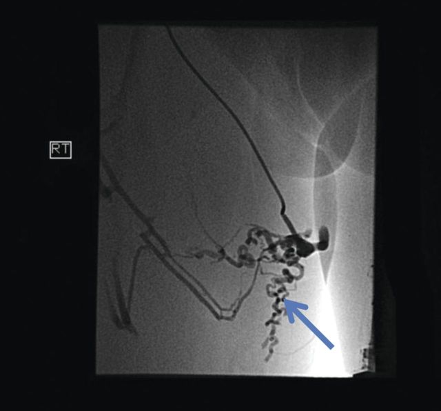
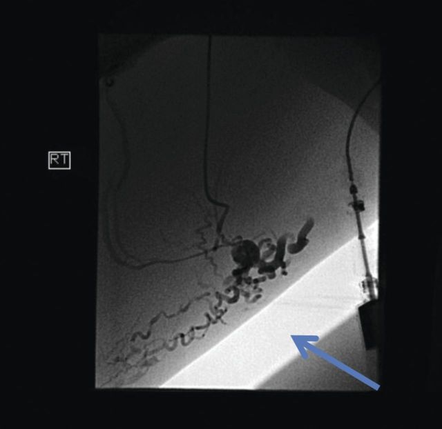
Varicose veins in the vulvar and peri-vulvar area are seen in 4% of women and most commonly seen during pregnancy. It is thought to be as a direct result of the presence of ovarian and pelvic varicosities. Diagnostic modalities used in the investigation of this condition included pelvic ultrasound, computed venography, magnetic resonance venography and catheter-directed venography. The treatment options in the past were hysterectomy and/or ligation of ovarian veins by open or laparoscopic approach. Modern techniques involve embolization of the varicosity via radiological techniques. In this case the patient presented with vulval and upper thigh varices associated with pelvic pain. They were located to be from the superficial external pudendal vein, which is not a common source but worth considering with other causes. They were treated successfully with fluoroscopy-guided embolization.
4%的女性会出现外阴及外阴周围区域的静脉曲张,且在孕期最为常见。这被认为是卵巢和盆腔静脉曲张直接导致的结果。用于该病症检查的诊断方法包括盆腔超声、计算机静脉造影、磁共振静脉造影和导管导向静脉造影。过去的治疗选择是通过开放手术或腹腔镜手术进行子宫切除术和/或卵巢静脉结扎。现代技术则是通过放射技术对静脉曲张进行栓塞。在该病例中,患者出现了与盆腔疼痛相关的外阴和大腿上部静脉曲张。经定位发现其源于浅表的阴部外静脉,这并非常见的来源,但与其他病因一同考虑时值得关注。通过荧光透视引导下的栓塞治疗,患者取得了成功治愈。