Kehoe Oksana, Cartwright Alison, Askari Ayman, El Haj Alicia J, Middleton Jim
Keele University, ISTM at RJAH Orthopaedic Hospital, Oswestry SY10 7AG, Shropshire, UK.
J Transl Med. 2014 Jun 3;12:157. doi: 10.1186/1479-5876-12-157.
Rheumatoid arthritis (RA) is a debilitating and painful disease leading to increased morbidity and mortality and novel therapeutic approaches are needed. The purpose of this study was to elucidate if mesenchymal stem cells (MSCs) injected in the joints of mice with arthritis are therapeutic, reducing joint swelling and cartilage destruction.
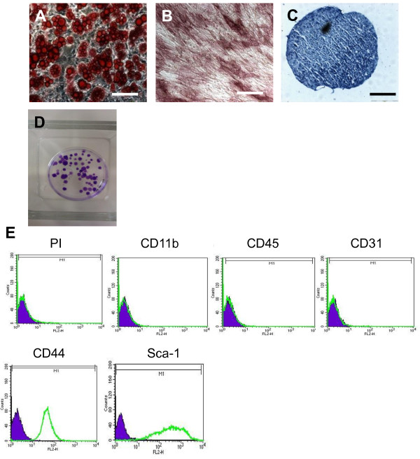
Murine mesenchymal stem cells (mMSCs) were isolated from bone marrow of C57Bl/6 mice and expanded in culture. Cells were tested for immunophenotype and their ability to form colonies and to differentiate into chondrocytes, osteocytes and adipocytes. Antigen-induced arthritis (AIA) was induced by intra-articular injection of methylated bovine serum albumin into the knee joints of preimmunized C57Bl/6 mice. After one day, when peak swelling occurs, 500,000 mMSCs labelled with red fluorescent cell tracker CM-DiI were injected intra-articularly in the right knee joint. Left knee joints were treated as controls by receiving PBS injections. Differences between groups were calculated by Mann Whitney U test or unpaired t tests using GraphPad Prism software version 5.
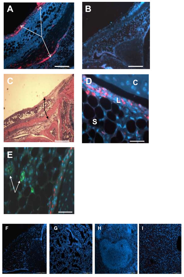
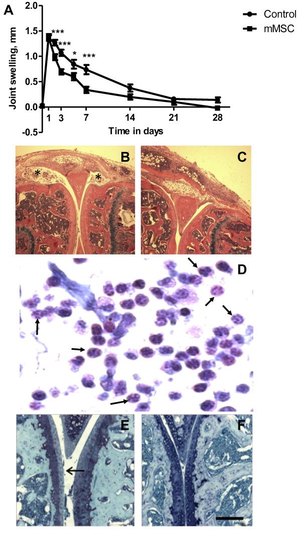
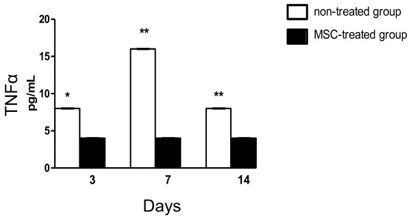
Knee joint diameter (swelling) was measured as a clinical indication of joint inflammation and this parameter was significantly less in MSC-treated mice compared to control-treated animals 48 hours after arthritis induction. This difference continued for ~7 days. CM-DiI-labelled MSCs were clearly visualised in the lining and sublining layers of synovium, in the region of the patella and femoral and tibial surfaces. By day 3, parameters indicative of disease severity, including cartilage depletion, inflammatory exudate and arthritic index were shown to be significantly reduced in MSC-treated animals. This difference continued for 7 days and was further confirmed by histological analysis. The serum concentration of tumour necrosis factor α was significantly decreased following MSC administration.
Our results reveal that MSCs injected in the joints of mice with AIA are therapeutic, reducing inflammation, joint swelling and cartilage destruction. These cells also integrate into the synovium in AIA.
类风湿性关节炎(RA)是一种使人衰弱且疼痛的疾病,会导致发病率和死亡率上升,因此需要新的治疗方法。本研究的目的是阐明注射到患有关节炎的小鼠关节中的间充质干细胞(MSC)是否具有治疗作用,能否减轻关节肿胀和软骨破坏。
从小鼠(C57Bl/6)骨髓中分离出小鼠间充质干细胞(mMSC)并在培养中进行扩增。检测细胞的免疫表型、形成集落的能力以及分化为软骨细胞、骨细胞和脂肪细胞的能力。通过向预先免疫的C57Bl/6小鼠膝关节内注射甲基化牛血清白蛋白诱导抗原诱导性关节炎(AIA)。一天后,当肿胀达到峰值时,将50万个用红色荧光细胞示踪剂CM-DiI标记的mMSC关节内注射到右膝关节中。左膝关节通过注射PBS作为对照。使用GraphPad Prism 5软件通过Mann Whitney U检验或非配对t检验计算组间差异。
测量膝关节直径(肿胀)作为关节炎症的临床指标,与对照处理的动物相比,在关节炎诱导后48小时,MSC处理的小鼠中该参数明显更低。这种差异持续约7天。在滑膜的衬里层和亚衬里层、髌骨区域以及股骨和胫骨表面区域可清晰看到CM-DiI标记的MSC。到第3天,在MSC处理的动物中,包括软骨消耗、炎性渗出物和关节炎指数在内的疾病严重程度指标显示显著降低。这种差异持续7天,并通过组织学分析进一步得到证实。给予MSC后,肿瘤坏死因子α的血清浓度显著降低。
我们的结果表明,注射到患有AIA的小鼠关节中的MSC具有治疗作用,可减轻炎症、关节肿胀和软骨破坏。这些细胞也会整合到AIA的滑膜中。