Roggia Murilo Felix, Imai Hirotaka, Shiraya Tomoyasu, Noda Yasuo, Ueta Takashi
Department of Ophthalmology, Graduate School of Medicine and Faculty of Medicine, The University of Tokyo, Tokyo, Japan.
School of Pharmaceutical Sciences, Kitasato University, Tokyo, Japan.
PLoS One. 2014 Jun 4;9(6):e98864. doi: 10.1371/journal.pone.0098864. eCollection 2014.
To evaluate the influence of glutathione peroxidase 4 (GPx4) expression in retinal pigment epithelium (RPE)/choroid tissue using a mouse model of laser-induced choroidal neovascularization (CNV).
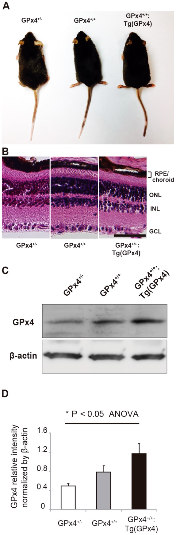
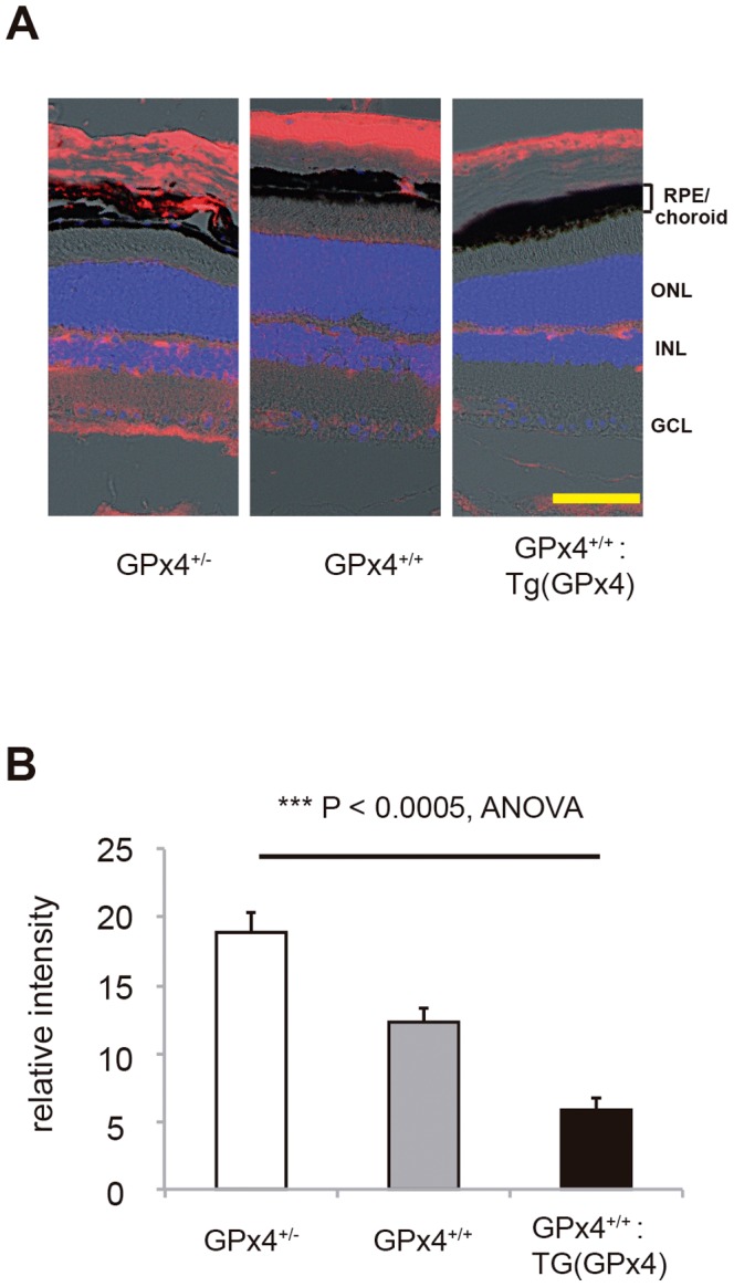
In this study, GPx4+/-, GPx4+/+, and GPx4-overexpressing transgenic mice were created for comparison. The mRNA and protein expression of vascular endothelial growth factor (VEGF)-A in RPE/choroid tissue were evaluated before and after CNV induction by laser. Moreover, we investigated the changes in the VEGF-A mRNA level in RPE/choroid tissue in the CNV model that have not been clearly shown previously. Lipid peroxidation in RPE/choroid tissue was evaluated by immunohistochemistry using antibody against 4-hydroxy-2-nonenal. To investigate the protective role of GPx4, the size of laser-induced CNV was compared on day 7 among the mice expressing different levels of GPx4.
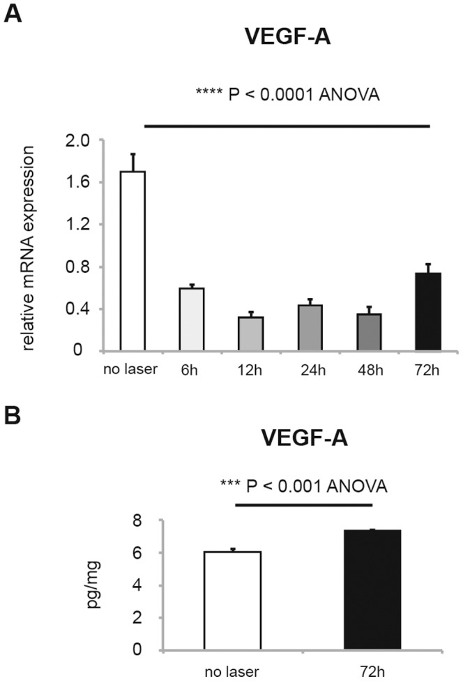
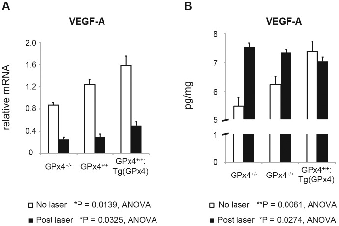
In the laser-induced CNV mouse model, laser treatment reduced the VEGF-A mRNA level in RPE/choroid tissue, while it increased the VEGF-A protein level. Evaluation of VEGF-A expression in RPE/choroid tissue of the GPx4+/-, GPx4+/+, and GPx4 transgenic mice revealed that GPx4 increased the VEGF-A protein level under physiological conditions (i.e., without laser treatment), while GPx4 suppressed the increase in the VEGF-A protein level under pathological conditions (i.e., after CNV induction by laser). In addition, GPx4 reduced the CNV size in a dose-dependent manner in vivo.
GPx4 suppresses the increase in the VEGF-A protein level, which occurs during the development of pathological CNV, thus partly explaining the protective effect of GPx4 against CNV.
利用激光诱导脉络膜新生血管(CNV)的小鼠模型,评估谷胱甘肽过氧化物酶4(GPx4)在视网膜色素上皮(RPE)/脉络膜组织中的表达影响。
在本研究中,创建了GPx4+/-、GPx4+/+和GPx4过表达转基因小鼠用于比较。通过激光诱导CNV前后,评估RPE/脉络膜组织中血管内皮生长因子(VEGF)-A的mRNA和蛋白表达。此外,我们研究了此前未明确显示的CNV模型中RPE/脉络膜组织中VEGF-A mRNA水平的变化。使用抗4-羟基-2-壬烯醛抗体通过免疫组织化学评估RPE/脉络膜组织中的脂质过氧化。为了研究GPx4的保护作用,比较了在第7天不同GPx4表达水平小鼠中激光诱导的CNV大小。
在激光诱导的CNV小鼠模型中,激光治疗降低了RPE/脉络膜组织中VEGF-A mRNA水平,同时增加了VEGF-A蛋白水平。对GPx4+/-、GPx4+/+和GPx4转基因小鼠的RPE/脉络膜组织中VEGF-A表达的评估显示,在生理条件下(即未进行激光治疗),GPx4增加了VEGF-A蛋白水平,而在病理条件下(即激光诱导CNV后),GPx4抑制了VEGF-A蛋白水平的增加。此外,GPx4在体内以剂量依赖方式减小了CNV大小。
GPx4抑制了病理性CNV发展过程中出现的VEGF-A蛋白水平升高,从而部分解释了GPx4对CNV的保护作用。