Olivares María José, González-Jamett Arlek M, Guerra María José, Baez-Matus Ximena, Haro-Acuña Valentina, Martínez-Quiles Narcisa, Cárdenas Ana M
Centro Interdisciplinario de Neurociencia de Valparaíso, Facultad de Ciencias, Universidad de Valparaíso, Playa Ancha, Valparaíso, Chile.
Departamento de Microbiología (Inmunología), Facultad de Medicina, Universidad Complutense de Madrid, Madrid, Spain.
PLoS One. 2014 Jun 5;9(6):e99001. doi: 10.1371/journal.pone.0099001. eCollection 2014.
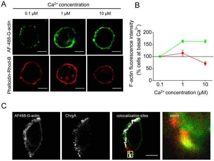
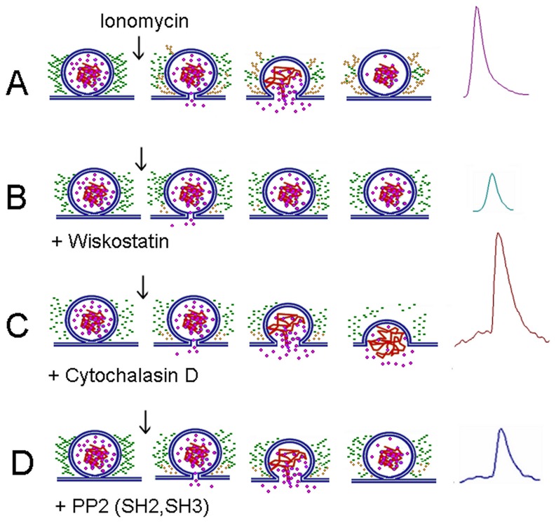
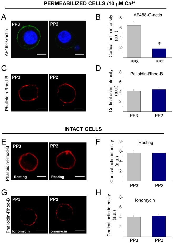
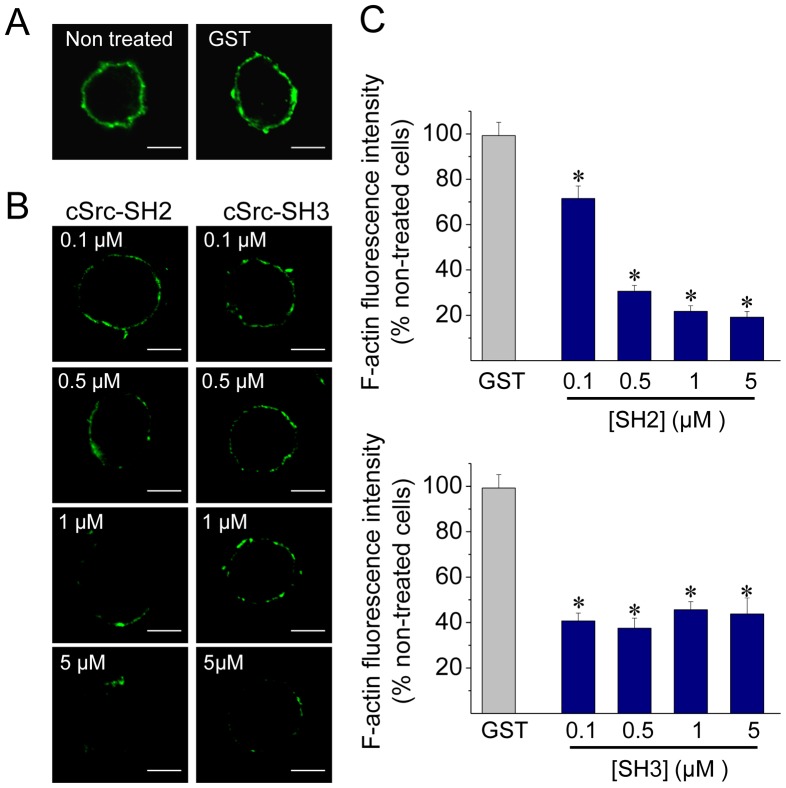
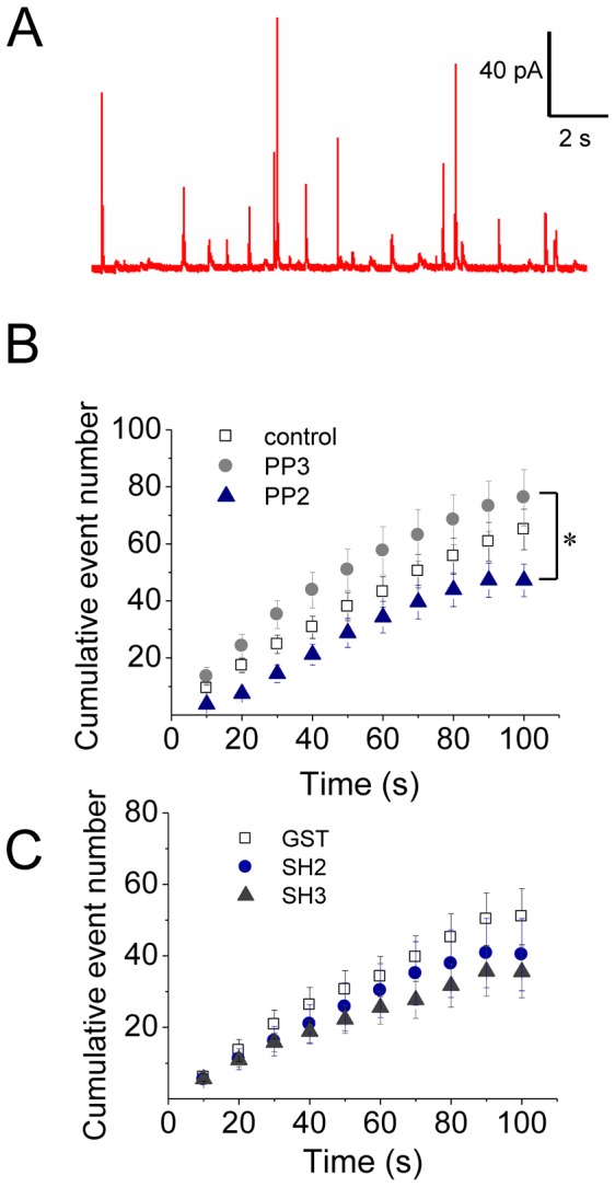
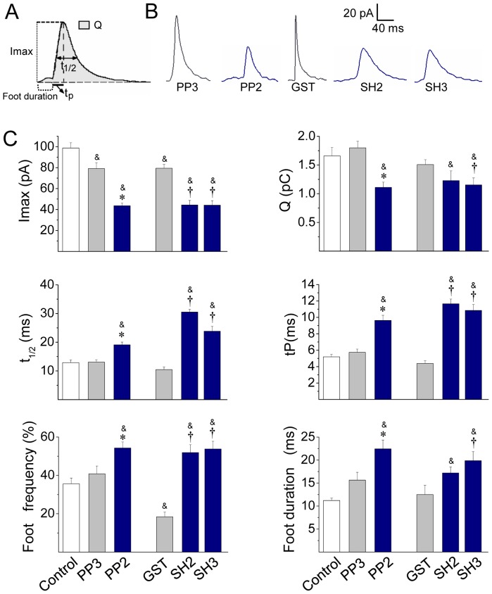
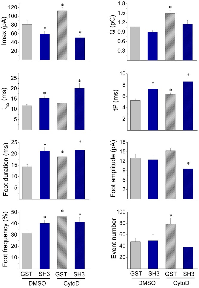
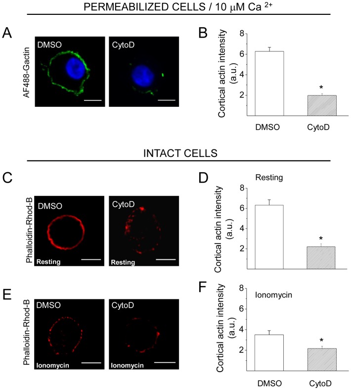
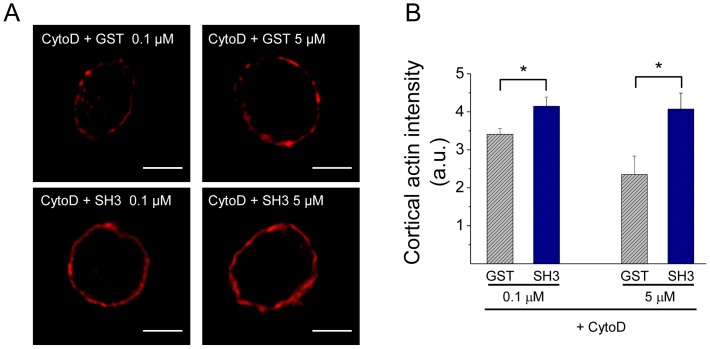
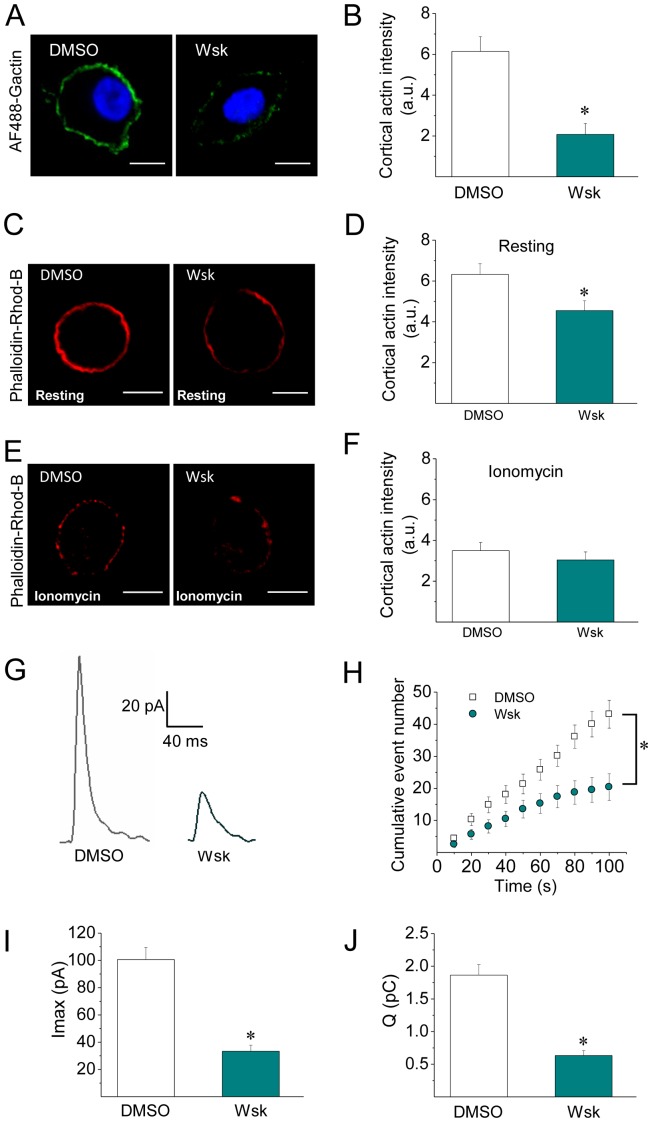
The cortical actin network is dynamically rearranged during secretory processes. Nevertheless, it is unclear how de novo actin polymerization and the disruption of the preexisting actin network control transmitter release. Here we show that in bovine adrenal chromaffin cells, both formation of new actin filaments and disruption of the preexisting cortical actin network are induced by Ca2+ concentrations that trigger exocytosis. These two processes appear to regulate different stages of exocytosis; whereas the inhibition of actin polymerization with the N-WASP inhibitor wiskostatin restricts fusion pore expansion, thus limiting the release of transmitters, the disruption of the cortical actin network with cytochalasin D increases the amount of transmitter released per event. Further, the Src kinase inhibitor PP2, and cSrc SH2 and SH3 domains also suppress Ca2+-dependent actin polymerization, and slow down fusion pore expansion without disturbing the cortical F-actin organization. Finally, the isolated SH3 domain of c-Src prevents both the disruption of the actin network and the increase in the quantal release induced by cytochalasin D. These findings support a model where a rise in the cytosolic Ca2+ triggers actin polymerization through a mechanism that involves Src kinases. The newly formed actin filaments would speed up the expansion of the initial fusion pore, whereas the preexisting actin network might control a different step of the exocytosis process.
在分泌过程中,皮质肌动蛋白网络会动态重排。然而,尚不清楚肌动蛋白的从头聚合以及先前存在的肌动蛋白网络的破坏是如何控制递质释放的。在此我们表明,在牛肾上腺嗜铬细胞中,触发胞吐作用的Ca2+浓度会诱导新肌动蛋白丝的形成以及先前存在的皮质肌动蛋白网络的破坏。这两个过程似乎调控胞吐作用的不同阶段;用N-WASP抑制剂威斯科他汀抑制肌动蛋白聚合会限制融合孔的扩张,从而限制递质的释放,而用细胞松弛素D破坏皮质肌动蛋白网络会增加每次事件释放的递质量。此外,Src激酶抑制剂PP2以及cSrc的SH2和SH3结构域也会抑制Ca2+依赖性肌动蛋白聚合,并减缓融合孔的扩张,而不会干扰皮质F-肌动蛋白的组织。最后,c-Src的分离的SH3结构域既能防止肌动蛋白网络的破坏,又能防止细胞松弛素D诱导的量子释放增加。这些发现支持了一种模型,即胞质Ca2+的升高通过一种涉及Src激酶的机制触发肌动蛋白聚合。新形成的肌动蛋白丝会加速初始融合孔的扩张,而先前存在的肌动蛋白网络可能控制胞吐作用过程的不同步骤。